Top
Published in: Journal of Gastrointestinal Cancer 1/2017

01-03-2017 | Case Report

Bile Duct Hamartoma Mimicking Metastatic Cholangiocarcinoma

Authors: Raymond Elsoueidi, Stephen J. Mularz, Elie M. Richa

Published in: Journal of Gastrointestinal Cancer | Issue 1/2017

Login to get access

Excerpt

A 63-year-old female presented to her primary care physician with weight loss of 30 lbs over the past 3 months, poor appetite, and jaundice. Her blood work revealed a total bilirubin of 12 mg/dl (range 0–1) with direct bilirubin of 10 mg/dl (range 0–0.3), alkaline phosphatase of 553 U/l (range 25–100), ALT 57 U/l (range 15–37), and AST 72 U/l (range 30–65). Further workup was performed including a CA 19–9 level of 549 U/ml (range 0–55) and magnetic resonance cholagiopancreatography (MRCP) revealing bile duct dilatation and multiple liver lesions concerning for metastatic disease. Computed tomography (CT) scan of the abdomen and pelvis showed multiple abnormally enhancing areas in the liver, the largest measuring 2.5 × 4.7 cm with dilated intra and extrahepatic bile ducts. She then had an endoscopic retrograde cholangiopancreatography (ERCP) that showed moderate dilation of the main bile duct as well as left and right hepatic ducts and all the intrahepatic branches along with distal common bile duct stenosis (Fig. 1). Bile duct brushing and biopsy were obtained. Atypical cells were found but were not consistent with malignancy. A biliary stent was placed. An endoscopic-guided ultrasound with fine needle aspiration of the head of pancreas was then performed and showed atypical ductal groups suspicious for adenocarcinoma. The patient underwent a laparoscopic evaluation with liver biopsy of the suspicious lesions and the pathology was consistent with bile duct hamartoma and no evidence of malignancy (Fig. 2). Her obstructive jaundice was due to common bile duct stenosis from benign fibrosis. The bilirubin and CA19-9 gradually decreased to 0.33 mg/dl and 90 U/ml, respectively. Her appetite improved and she started gaining weight. She is having her biliary stent changed every 3 months and CT scans of abdomen every 6 months with stable findings for the last 18 months.
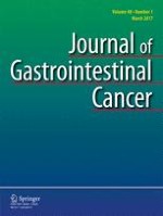
Literature
1.
go back to reference Wetter LA, Ring EJ, Pellegrini CA, Way LW. Differential diagnosis of sclerosing cholangiocarcinomas of the common hepatic duct (Klatskin tumors). Am J Surg J. 1991;161(1):57–62.CrossRef Wetter LA, Ring EJ, Pellegrini CA, Way LW. Differential diagnosis of sclerosing cholangiocarcinomas of the common hepatic duct (Klatskin tumors). Am J Surg J. 1991;161(1):57–62.CrossRef
2.
go back to reference Verbeek PC, van Leeuwen DJ, de Wit LT, et al. Benign fibrosing disease at the hepatic confluence mimicking Klatskin tumors. Surgery. 1992;112(5):866–71.PubMed Verbeek PC, van Leeuwen DJ, de Wit LT, et al. Benign fibrosing disease at the hepatic confluence mimicking Klatskin tumors. Surgery. 1992;112(5):866–71.PubMed
3.
go back to reference Venkatanarasimha N, Thomas R, Armstrong EM, et al. Imaging features of ductal plate malformations in adults. Clin Radiol. 2011;66:1086–109.CrossRefPubMed Venkatanarasimha N, Thomas R, Armstrong EM, et al. Imaging features of ductal plate malformations in adults. Clin Radiol. 2011;66:1086–109.CrossRefPubMed
5.
go back to reference Terada T, Moriki T. Monolobar hepatobiliary fibropolycystic disease. Pathol Oncol Res. 2011;7:159–65.CrossRef Terada T, Moriki T. Monolobar hepatobiliary fibropolycystic disease. Pathol Oncol Res. 2011;7:159–65.CrossRef
6.
go back to reference Desmet VJ. Congenital diseases of intrahepatic bile ducts: variations on the theme “ductal plate malformation”. Hepatology. 1992;16:1069–83.CrossRefPubMed Desmet VJ. Congenital diseases of intrahepatic bile ducts: variations on the theme “ductal plate malformation”. Hepatology. 1992;16:1069–83.CrossRefPubMed
8.
go back to reference Semelka RC, Hussain SM, Marcos HB, et al. Biliary hamartomas: solitary and multiple lesions shown on current MR techniques including gadolinium enhancement. J Magn Reson Imaging. 1999;10:196–201.CrossRefPubMed Semelka RC, Hussain SM, Marcos HB, et al. Biliary hamartomas: solitary and multiple lesions shown on current MR techniques including gadolinium enhancement. J Magn Reson Imaging. 1999;10:196–201.CrossRefPubMed
9.
go back to reference Nagano Y, Matsuo K, Gorai K, et al. Bile duct hamartomas (von Meyenburg complexes) mimicking liver metastasis from bile duct cancer: MRC findings. World J Gastroenterol. 2006;12:1321–3.CrossRefPubMedPubMedCentral Nagano Y, Matsuo K, Gorai K, et al. Bile duct hamartomas (von Meyenburg complexes) mimicking liver metastasis from bile duct cancer: MRC findings. World J Gastroenterol. 2006;12:1321–3.CrossRefPubMedPubMedCentral
11.
go back to reference Rocken C, Pross M, Brucks U, et al. Cholangiocarcinoma occurring in a liver with multiple bile-duct hamartomas (von Meyenburg complexes). Arch Pathol Lab Med. 2000;124:1704–6.PubMed Rocken C, Pross M, Brucks U, et al. Cholangiocarcinoma occurring in a liver with multiple bile-duct hamartomas (von Meyenburg complexes). Arch Pathol Lab Med. 2000;124:1704–6.PubMed
Metadata
Title
Bile Duct Hamartoma Mimicking Metastatic Cholangiocarcinoma
Authors
Raymond Elsoueidi
Stephen J. Mularz
Elie M. Richa
Publication date
01-03-2017
Publisher
Springer US
Published in
Journal of Gastrointestinal Cancer / Issue 1/2017
Print ISSN: 1941-6628
Electronic ISSN: 1941-6636
DOI
https://doi.org/10.1007/s12029-016-9802-z

Other articles of this Issue 1/2017

Journal of Gastrointestinal Cancer 1/2017 Go to the issue