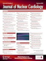
A 47-year-old man presented with progressive precordial discomfort and breathlessness over 10 days, without fever, nausea, night sweats, or weight loss. Two days before admission, bedside echocardiography detected signs of tamponade in local medical center. Pericardiocentesis was performed and blood-stained flood was drained but symptoms didn’t improve. Transesophageal echocardiography revealed a huge (69 × 37 mm) right atrial mass (Panel A), which is consistent with cardiovascular-enhanced CT (Panel B, Supplementary material, video S1), also showing that the huge right atrial mass infiltrated the pericardium. Contrast-enhanced echocardiography showed vascularity (Panel C). Fluorodeoxyglucose positron emission tomography demonstrated tracer uptake and multiple bone metastases (Panel D, Supplementary material, Video S2, S3 and S4). Bone marrow biopsy and immunohistochemistry test were performed, which showed spindle cell existence (Supplementary material, Panel E) and positive of CD31, CD34, and ERG, suggesting a diagnosis of primary cardiac angiosarcoma.