


Published in:
Open Access 01-12-2013 | Imaging in Cardiology
Living with high output
Published in: Netherlands Heart Journal | Issue 12/2013
Login to get accessExcerpt
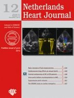
A 59-year-old woman presented for evaluation of heart failure. As a child she was known to have a ventricular septum defect which was no longer demonstrated during adulthood. She was normally in good shape but experienced yearly episodes of left-sided lower airway infections. During the last episode her general physician heard an ejection murmur and rales over her lungs. Chest radiography revealed an enlarged heart, a right descending aorta and a prominent left pulmonary artery (Fig. 1). There were no signs of congestion. Echocardiography revealed a slightly dilated left ventricle with an ejection fraction of 43%. Magnetic resonance imaging showed an anomalous left pulmonary artery originating from the right descending aorta (Fig. 2). The heart also appeared to be enlarged and the ejection fraction was slightly higher at 52%. Moreover, a high cardiac output of 11.6 l per minute was measured.
Fig. 1
Chest radiography showing an enlarged heart figure, a right descending aorta (white arrow) and an enlarged left pulmonary artery (black arrow)
Fig. 2
Magnetic resonance imaging showing the left pulmonary artery originating from the ascending aorta (arrow)
× ![]()
× ![]()
…