


Published in:
01-04-2015 | Symposium: 2014 Bernese Hip Symposium
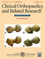
Biochemical MRI Predicts Hip Osteoarthritis in an Experimental Ovine Femoroacetabular Impingement Model
Published in: Clinical Orthopaedics and Related Research® | Issue 4/2015
Login to get accessAbstract
Background
Cam-type femoroacetabular impingement (FAI) resulting from an abnormal nonspherical femoral head shape leads to chondrolabral damage and is considered a cause of early osteoarthritis. A previously developed experimental ovine FAI model induces a cam-type impingement that results in localized chondrolabral damage, replicating the patterns found in the human hip. Biochemical MRI modalities such as T2 and T2* may allow for evaluation of the cartilage biochemistry long before cartilage loss occurs and, for that reason, may be a worthwhile avenue of inquiry.
Questions/purposes
We asked: (1) Does the histological grading of degenerated cartilage correlate with T2 or T2* values in this ovine FAI model? (2) How accurately can zones of degenerated cartilage be predicted with T2 or T2* MRI in this model?
Methods
A cam-type FAI was induced in eight Swiss alpine sheep by performing a closing wedge intertrochanteric varus osteotomy. After ambulation of 10 to 14 weeks, the sheep were euthanized and a 3-T MRI of the hip was performed. T2 and T2* values were measured at six locations on the acetabulum and compared with the histological damage pattern using the Mankin score. This is an established histological scoring system to quantify cartilage degeneration. Both T2 and T2* values are determined by cartilage water content and its collagen fiber network. Of those, the T2* mapping is a more modern sequence with technical advantages (eg, shorter acquisition time). Correlation of the Mankin score and the T2 and T2* values, respectively, was evaluated using the Spearman’s rank correlation coefficient. We used a hierarchical cluster analysis to calculate the positive and negative predictive values of T2 and T2* to predict advanced cartilage degeneration (Mankin ≥ 3).
Results
We found a negative correlation between the Mankin score and both the T2 (p < 0.001, r = −0.79) and T2* values (p < 0.001, r = −0.90). For the T2 MRI technique, we found a positive predictive value of 100% (95% confidence interval [CI], 79%–100%) and a negative predictive value of 84% (95% CI, 67%–95%). For the T2* technique, we found a positive predictive value of 100% (95% CI, 79%–100%) and a negative predictive value of 94% (95% CI, 79%–99%).
Conclusions
T2 and T2* MRI modalities can reliably detect early cartilage degeneration in the experimental ovine FAI model.
Clinical Relevance
T2 and T2* MRI modalities have the potential to allow for monitoring the natural course of osteoarthrosis noninvasively and to evaluate the results of surgical treatments targeted to joint preservation.