


Published in:
01-02-2017 | Stanford Multidisciplinary Seminars
Radiofrequency Ablation of Cameron Lesions
Published in: Digestive Diseases and Sciences | Issue 2/2017
Login to get accessExcerpt
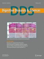
A 76-year-old woman was initially evaluated for weakness and recurrent melena accompanied by severe iron deficiency anemia. First diagnosed with iron deficiency anemia due to Cameron lesions 4 years prior to presentation, she was aggressively treated with oral and intravenous (IV) iron supplementation and omeprazole 40 mg twice daily. She had decided to forgo laparoscopic hernia repair and fundoplication fearful of potential perioperative complications. Her past medical history was otherwise significant for nonalcoholic steatohepatitis, hypertension, and bilateral pulmonary emboli that had resolved after the institution of IV heparin, IV tissue plasminogen activator, and oral warfarin therapy. Because she was heterozygous for a Leiden factor V mutation, an unfixed inferior vena cava filter had been placed. There was no history of aspirin or nonsteroidal anti-inflammatory drug (NSAID) use. On admission, she was pale, with hemoglobin of 8.1 mg/dl. An upper endoscopy demonstrated a large sliding hiatal hernia with several linear mucosal erosions at the level of the diaphragmatic hiatus without stigmata of recent hemorrhage (Fig. 1, left panel). Barium esophagram (Fig. 2, left panel) and computed tomography (CT) of the thorax (Fig. 2 middle and right panels) confirmed the large sliding hiatal hernia (HH). Because of the ongoing melena and anemia, she was treated with HALO®-90 radiofrequency ablation (RFA) (Barrx™ RF Ablation, Covidien GI Solutions, Sunnyvale, CA) applied circumferentially at the level of the diaphragmatic pinch (Fig. 1, middle and right panels) which stabilized her clinically for >2 weeks, after which she underwent hernia repair and partial fundoplication.
Fig. 1
Left panel Upper endoscopy revealing a large sliding hiatal hernia and several linear erosions at the diaphragmatic hiatus without stigmata of recent hemorrhage. Middle and right panels Endoscopic appearance immediately after HALO®-90 radiofrequency ablation delivered in a circumferential fashion at the level of the diaphragmatic pinch. The HALO®-90 probe is visible at the top of the images
Fig. 2
Left panel Barium esophagram revealing a large, sliding hiatal hernia. Middle and right panels Coronal CT scan views demonstrating the hiatal hernia. Cameron lesions are not discernible in either of these two imaging modalities
× ![]()
× ![]()
…