


Published in:
01-08-2017
Extraskeletal Osteosarcoma Arising from the Pleura
Published in: Lung | Issue 4/2017
Login to get accessExcerpt
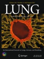
A 75-year-old woman presented at Emergency Department with acute onset of right-sided chest pain and hemoptysis. Radiological examination revealed a large thoracic mass located in the right upper lung field. Chest CT scan showed a large round-shaped tumor (82 × 81 mm), heterogeneously enhanced after the injection of iodine contrast (Fig. 1a–c) with no clear signs of chest wall invasion. At 18F-FDG PET/CT scan, the thoracic mass showed a moderate (SUVmax = 4.45), non-homogenous uptake of the tracer without further uptaking areas (Fig. 1d, e). Percutaneous needle biopsy showed malignant p63+, TTF1-cells, raising the suspicion of squamous cell carcinoma of the lung. Accordingly, a surgical excision was planned for right lateral thoracotomy. At surgery, the tumor appeared as a well-encapsulated, cystic and huge mass arising from parietal pleura without involvement of chest wall but strictly adherent to the visceral pleura and pulmonary parenchyma. Radical resection was achieved with wedge resection of right upper lobe, while chest wall resection was not needed.
Fig. 1
Radiological evaluation. Axial (a), sagittal (b), and frontal (c) CT scan showing a large round-shaped mass (arrows), with intra-lesional fluid non-contrast-enhanced areas (asterisk); Radiometabolic evaluation: 18F-FDG PET/CT scan revealed the moderate uptake of the tracer at this level (arrows in d and e)
× ![]()
…