


Published in:
01-06-2009 | Correspondence
Facial necrotizing fasciitis in an infant caused by a five toxin-secreting methicillin-susceptible Staphylococcus aureus
Published in: Intensive Care Medicine | Issue 6/2009
Login to get accessExcerpt
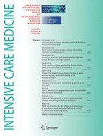
Sir: A 10-month-old boy returning from Egypt was admitted to the paediatric department for a 4-day history of fever and diarrhoea. He had a maculopapular rash and a chin furuncle with no history of trauma. A methicillin-susceptible Staphylococcus aureus (MSSA) was cultured from the pus. A first skin biopsy showed cellulitis with superficial necrosis that did not reach the underlying fascia. He received oxacillin, gentamicin, and diarrhoea treatment. After 72 h, he was transferred to the PICU for septic shock. He had a necrotic ulcer on the chin, conjunctivitis, and trunk desquamation (Fig. 1a). Blood tests showed thrombocytopenia, hyponatraemia, hyperlactataemia, liver cytolysis, and renal failure. He received fluids, vasopressors, endotracheal ventilation, analgesia, and four new antibiotics (cefotaxime, vancomycin, rifampin, and metronidazole). The skin lesion spread within 12 h and the oedema extended from the chin to the lower right ear. Because MRI was unavailable, cervical computed tomography was performed after clinical stabilization. Findings suggested necrotizing fasciitis (NF). Emergent surgical debridement showed extensive necrosis reaching the underlying muscles. A new skin biopsy performed during surgery confirmed the diagnosis (Fig. 1b). A new bacteriological sample identified the same MSSA, as shown by chromosomal DNA restriction patterns in pulsed-field gel electrophoresis. PCRs identified genes coding for Panton Valentine leukocidin (PVL); A, H, and K-enterotoxins; and toxic-shock-syndrome toxin-1 (TSST-1). The patient improved rapidly after surgery with drainage for 6 days (Fig. 1c). He was discharged home 3 weeks later with oral antibiotics for a total of 6 weeks. After 1 year, healing was complete with no skin graft (Fig. 1d).
Fig. 1
a Necrotic lesion of the chin and massive hemifacial oedema during resuscitation in the PICU immediately before surgery. Note the skin desquamation on the left shoulder. b The skin biopsy performed during surgery showed necrosis of skeletal and adipose tissues with diffuse pycnotic neutrophilic infiltration extending to the underlying fascia and muscle (haematoxylin–eosin stained section, ×400). c Immediate post-operative appearance of the lesion showing the drain that was inserted after excision of all necrotic tissues. d Patient 1 year later
× ![]()
…