


Published in:
01-08-2017 | Images that Teach
Incidental detection of abnormal 99mTc-sestamibi uptake in the sternum and ribcage from multiple myeloma by SPECT myocardial perfusion imaging
Published in: Journal of Nuclear Cardiology | Issue 4/2017
Login to get accessExcerpt
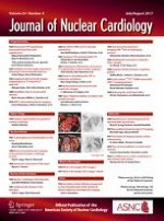
A 66-year-old man with a past medical history of coronary artery bypass grafting, diabetes mellitus, hypertension, heart failure, and nonalcoholic steatohepatitis underwent regadenoson myocardial perfusion imaging (MPI) using 99mTc-sestamibi for evaluation of shortness of breath. Stress and rest ECGs were unremarkable. The images showed decreased perfusion in the inferolateral wall with no change from stress to rest consistent with a moderate-sized scar in the distribution of left circumflex coronary artery involving 15% of LV myocardium (Figure 1a). The left ventricular ejection fraction was 52%. Markedly abnormal radiotracer uptake in the sternum and the rib cage is noted on the tomographic (arrows in Figure 1a) and the rotating raw images (arrows in Figure 1b). The patient underwent bone marrow biopsy which confirmed the presence of multiple myeloma and subsequently was referred for bone marrow transplantation.
Figure 1
A Stress/rest SPECT myocardial perfusion images show fixed scar in the inferolateral wall involving 15% of LV myocardium. In addition, there is abnormal tracer uptake in the ribs which are in close proximity to the heart (yellow arrows). B Planar oblique chest images show increased tracer uptake in the sternum and ribs (white arrows)
× ![]()
…