


Published in:
01-12-2016 | Imaging in Intensive Care Medicine
Common femoral artery pseudoaneurysm
Published in: Intensive Care Medicine | Issue 12/2016
Login to get accessExcerpt
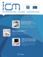
A 19-year-old female patient was admitted to the intensive care unit 15 days ago, having suffered a severe traumatic brain injury. On physical examination, a small bulging hematoma was observed on the right inguinal region, where several arterial blood samplings were performed during her hospitalization. Ultrasonographic (US) scanning showed an anechoic pulsating cavity (Fig. 1a) on superficial tissues, communicating with the common femoral artery (CFA) (Fig. 1b). Bidirectional flow was detected on color and spectral Doppler imaging (Fig. 1b, c) and a final diagnosis of CFA pseudoaneurysm was made. US-guided compression over 10 min successfully thrombosed the cavity and stopped the arterial leak (Fig. 1d). An arterial pseudoaneurysm (PSA) is a contained rupture occurring predominantly after arterial catheterization for diagnostic and interventional purposes, but also possible after needle-stick injuries and an inadequate compression after the procedure. The most catastrophic complication is rupture, most frequent with PSA larger than 3 cm. Common treatment options include US-guided compression, with an overall success rate of 75–98 %, US-guided thrombin injection (91–100 % success rate), and eventually surgical repair if previous treatments fail.
Fig. 1
a Right inguinal region ultrasonography (US) demonstrating a cavity (calipers) on superficial tissues, measuring 12.4 × 8.7 mm. CFA common femoral artery. b Color Doppler demonstrating the communication of the cavity with the common femoral artery and bidirectional flow (blue and red color within the cavity, also called the “ying-yang” sign). Arrows pseudoaneurysm neck. c Pulsed-wave Doppler showing a high-velocity bidirectional flow. d After US-guided compression, the cavity is thrombosed (asterisk) and flow was no longer detected. CFA common femoral artery
× ![]()
…