


Published in:
01-01-2020 | Image–Obstetrics & Gynecology
Visualization of intrahepatic portosystemic shunt and intra-abdominal umbilical vein varix using fetal ultrasonography with HDlive flow imaging
Published in: Journal of Medical Ultrasonics | Issue 1/2020
Login to get accessExcerpt
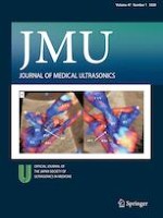
A 39-year-old Japanese multiparous woman presented to our hospital at 25 weeks of gestation for evaluation of fetal anomalies. On examining her using fetal ultrasonography (Voluson E10; GE Healthcare Japan, Tokyo, Japan), we observed fetal intra-abdominal umbilical vein varix (FIUVV), with the maximum diameter of the vein being 11 mm. At 28 weeks of gestation, an aneurysmal vascular enlargement in the right liver was first detected. In addition to conventional two-dimensional (2D) ultrasound imaging, three-dimensional (3D) HDlive flow imaging revealed that the blood flow of the portal vein was directly associated with the bifurcated hepatic vein through the intra-right liver aneurysmal lesioned portion (Fig. 1a, b). Based on these findings, we diagnosed the vascular anomaly as congenital intrahepatic portosystemic shunt (IPSS). Amniotic fluid chromosomal examination confirmed trisomy 21 in the fetus. At 35 + 5 weeks of gestation, an emergent cesarean section was performed due to a non-reassuring fetal status. A 1763-g female infant was delivered with an Apgar score of 8 and 9 at 1 and 5 min, respectively. Postnatal 2D ultrasound and CT scan indicated IPSS, similar to the prenatal diagnosis (Fig. 1c). The presence of a bifurcated hepatic vein was confirmed postnatally and was considered a vascular anomaly. During the neonatal period, no significant elevation in the galactose, total bile acid, or ammonia levels was determined on serial blood tests.
Fig. 1
a Three-dimensional HDlive flow imaging of the IPSS and FIUVV using fetal ultrasonography. Visualization from the right side of the fetus (left image) and the left side of the fetus (right image). The portal vein is connected to the bifurcated hepatic vein through the aneurysmal portion (*) in the liver. b Two-dimensional ultrasonographic findings of the fetal thoracicoabdominal region. The diameter of the UVV is about 12 mm. The asterisk shows the aneurysmal portion in the liver, which is a finding of IPSS. c Postnatal computed tomography (CT) examination image, which shows the portal venous phase (colored green) of a contrast CT scan on postnatal day 48. The asterisk shows the IPSS. LMB shows the prenatally detected portal vein connecting the portal sinus and aneurysmal portion in the liver. HV shows the prenatally detected hepatic vein between the aneurysmal portion in the liver and IVC. RASB is another portal vein connected to the aneurysmal portion, which was not detected prenatally. PV portal vein, UVV umbilical vein varix, HV hepatic vein, CA celiac artery, IVC inferior vena cava, DV ductus venosus, RA right atrium, dAo descending aortic artery, PS portal sinus, RASB right anterior superior branch of the portal vein, LMB left medial branch of the portal vein, MPV main portal vein, IPSS intrahepatic portosystemic shunt, FIUVV fetal intra-abdominal umbilical vein varix
× ![]()
…