Top
Published in: Dysphagia 2/2014

01-04-2014 | Clinical Conundrum

Unusual Cause for Smoldering Dysphagia

Authors: Pedro C. Figueiredo, Pedro Pinto-Marques, Paula Borralho, João Freitas

Published in: Dysphagia | Issue 2/2014

Login to get access

Excerpt

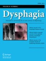
A 30-year-old black woman presented with heartburn and odynophagia. She had a 2-year history of Behçet’s disease and systemic lupus erythematosus and had been treated with colchicine, hydroxychloroquine, and sucralfate. Odynophagia was not related to the presence of oral ulcers as they were painless and when they were in remission the patient would still intermittently complain of substernal pain. The patient underwent upper digestive endoscopy that revealed only small mucosal irregularities in the upper third of the esophagus (Fig. 1). Biopsies of these segments showed marked acanthosis and papillomatosis of the squamous epithelium as well as intense lymphoplasmacytic infiltrate with an increased number of intraepithelial lymphocytes (IEL). There were neither granulocytes nor signs of viral infection. The endoscopic findings were then attributed to regenerative changes of the epithelium and the patient was started on a proton pump inhibitor (PPI), assuming gastroesophageal reflux disease (GERD). During the following years there were flare-ups of rheumatologic disease activity due to the patient’s lack of adherence to therapy. However, there was no correlation of the patient’s maintained (although scarce) complaints of transitory dysphagia and substernal pain.
Literature
1.
go back to reference Rubio CA, Sjodahl K, Lagergren J. Lymphocytic esophagitis: a histologic subset of chronic esophagitis. Am J Clin Pathol. 2006;125:432–7.PubMedCrossRef Rubio CA, Sjodahl K, Lagergren J. Lymphocytic esophagitis: a histologic subset of chronic esophagitis. Am J Clin Pathol. 2006;125:432–7.PubMedCrossRef
2.
go back to reference Hori K, Watari J, Fukui H, et al. Do endoscopic features suggesting eosinophilic esophagitis represent histological eosinophilia? Dig Endosc. 2013. doi:10.1111/den.12091.PubMed Hori K, Watari J, Fukui H, et al. Do endoscopic features suggesting eosinophilic esophagitis represent histological eosinophilia? Dig Endosc. 2013. doi:10.​1111/​den.​12091.PubMed
4.
go back to reference Yi SW, Cheon JH, Kim JH, et al. The prevalence and clinical characteristics of esophageal involvement in patients with Behçet’s disease: a single center experience in Korea. J Korean Med Sci. 2009;24:52–6.PubMedCentralPubMedCrossRef Yi SW, Cheon JH, Kim JH, et al. The prevalence and clinical characteristics of esophageal involvement in patients with Behçet’s disease: a single center experience in Korea. J Korean Med Sci. 2009;24:52–6.PubMedCentralPubMedCrossRef
Metadata
Title
Unusual Cause for Smoldering Dysphagia
Authors
Pedro C. Figueiredo
Pedro Pinto-Marques
Paula Borralho
João Freitas
Publication date
01-04-2014
Publisher
Springer US
Published in
Dysphagia / Issue 2/2014
Print ISSN: 0179-051X
Electronic ISSN: 1432-0460
DOI
https://doi.org/10.1007/s00455-013-9489-2

Other articles of this Issue 2/2014

Dysphagia 2/2014 Go to the issue