


Published in:
01-07-2018 | Images in Anesthesia
Nasogastric tube location: a diagnostic dilemma
Published in: Canadian Journal of Anesthesia/Journal canadien d'anesthésie | Issue 7/2018
Login to get accessExcerpt
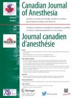
A 79-yr-old woman was admitted to hospital with presumed respiratory sepsis. Her past medical history included hypertension, coronary artery disease, and a hiatus hernia. She was initially managed with non-invasive ventilation, antibiotics, and vasopressors. To allow enteral feeding, a wide-bore (14 Fr) nasogastric tube (NGT) was inserted, and its position was provisionally confirmed using auscultation of injected air, although nothing could be aspirated from it. Chest radiography (CXR) was performed for definitive confirmation of the NGT position before initiating feeding, which revealed that the NGT tip appeared to be in the right lung base (Figure). Assuming misplacement, the NGT was removed, and the procedure was abandoned. Later the same day, the patient’s respiratory status deteriorated, and she was intubated and ventilated.
Figure
A) Chest radiograph showing a nasogastric tube overlying the right lung field. The black arrow indicates the tip of the nasogastric tube that appears to be in the right hemi-thorax. B) Computed tomography image of the chest showing a large hiatal hernia (arrow) extending into the right hemi-thorax. The images were used with the patient’s permission
× ![]()
…