


Published in:
01-05-2010 | Correspondence
Hypercytokinemia with 2009 pandemic H1N1 (pH1N1) influenza successfully treated with polymyxin B-immobilized fiber column hemoperfusion
Published in: Intensive Care Medicine | Issue 5/2010
Login to get accessExcerpt
In September 2009, a 16-year-old female with no medical history developed fever, general fatigue, and diarrhea. A rapid diagnosis kit at a local clinic showed type A influenza. She was given 10 mg zanamivir inhalation, twice daily. The following day, her body temperature rose to 41.7°C and she experienced respiratory distress. She was transported to our hospital by ambulance. On arrival, her blood pressure was 90/40 mmHg, heart rate 150/min, and respiratory rate 35/min. She was administered a large dose of crystalloid fluid and norepinephrine (0.3 μg/kg/min). Mechanical ventilation with endotracheal intubation was begun. Purulent sputum removed by suction contained Gram-positive cocci on a Gram-stained smear. Cultures of blood, urine, and stool testing for various pathogens were negative. Mechanical ventilation was performed in pressure control mode with PaO2/FiO2 (P/F) ratio of 148. Thereafter, a recruitment maneuver was performed, switching to airway pressure release ventilation (APRV) mode. P/F ratio temporarily improved to 224, but then deteriorated to 165. Oseltamivir (150 mg/day), sivelestat sodium hydrate (300 mg/day), and antibiotics (initially ampicillin/sulbactam 6 g/day) were administered. On day 2, complicated by disseminated intravascular coagulation, no improvement in respiratory status was observed, prompting an increase in oseltamivir dosage to 300 mg/day. Type A influenza was confirmed as 2009 pandemic H1N1 (pH1N1) virus by polymerase chain reaction (PCR). On day 3, methicillin-resistant Staphylococcus aureus (MRSA) was identified in the sputum collected immediately after intubation. Antibiotics were changed to linezolid (12 g/day) plus clindamycin (1,800 mg/day) for MRSA. Serum cytokine levels were highly elevated (Fig. 1). We considered that the severe respiratory failure might be related to hypercytokinemia caused by pH1N1 and MRSA infection. With no improvement in oxygenation, polymyxin B-immobilized fiber column (PMX) hemoperfusion was begun in an attempt to reduce the inflammatory mediators and improve oxygenation. Oxygenation gradually improved; after 1 h, the P/F ratio increased from 144 to 184, and after 8 h to 308. PMX hemoperfusion was performed for 14 h; following cessation, oxygenation declined (P/F ratio 220). On days 4 and 5, PMX hemoperfusion was planned for 18 h on each day, and the P/F ratio increased to 407. Serum inflammatory mediators decreased to normal levels after the PMX treatments. She was extubated and discharged from the hospital.
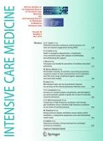
Fig. 1
Clinical course of the patient. PaO2/FiO2 (P/F) ratio and serum inflammatory cytokine levels improved after PMX hemoperfusion on three consecutive days. SBT/ABPC ampicillin/sulbactam, LZD linezolid, CLDM clindamycin, VCM vancomycin
× ![]()
…