


Published in:
01-04-2017 | Imaging in Intensive Care Medicine
Documenting the invisible in stroke-like symptoms during extracorporeal membrane oxygenation
Published in: Intensive Care Medicine | Issue 4/2017
Login to get accessExcerpt
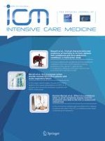
A 17-year-old male with dilated cardiomyopathy developed cardiac arrest during hospital admission. Awakening was achieved during veno-arterial extracorporeal membrane oxygenation (ECMO). Seven days after cardiac arrest, he developed lethargy, global aphasia, and right hemiplegia. Left hemispheric infarction was suspected. However, his brain computed tomography was unremarkable. Magnetic resonance imaging (MRI) of the brain was performed soon after weaning from ECMO. Routine MRI sequences also showed no specific findings (Fig. 1a–d). However, susceptibility-weighted imaging (SWI) revealed hundreds of small signal loss lesions (Fig. 1e). His stroke-like symptoms resolved over the course of a month, but the SWI lesions were still observable 1 year later (Fig. 1f).
Fig. 1
Diffusion-weighted imaging (a), T1-weighted imaging (b), T2-weighted imaging (c), and fluid attenuated inversion recovery imaging (d) were unremarkable 15 days after presentation. However, susceptibility-weighted imaging revealed hundreds of cerebral microbleeds (e). Cerebral microbleeds were still observable on follow-up susceptibility-weighted imaging performed 1 year later (f)
× ![]()
…