


Published in:
01-12-2016 | Images in Hematology
Chédiak–Higashi-like granules and waxy Auer bodies in a case of acute promyelocytic leukemia
Published in: International Journal of Hematology | Issue 6/2016
Login to get accessExcerpt
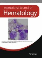
A 31-year-old man presented with pharyngeal pain and purpura on the right dorsal surface of the hand for 1 week. The complete blood count showed leukopenia and thrombocytopenia (white blood cell count, 1.8 × 10⁹/L; platelet count, 51 × 109/L). Coagulation tests revealed the following: prothrombin time–international normalized ratio (PT–INR) of 1.46; fibrinogen level of 36 mg/dL; fibrin degradation products (FDP) level of 50.9 μg/mL; D-dimer level of 60.6 μg/mL; and thrombin–antithrombin III (TAT) complex level of 54.6 ng/mL. These results indicated disseminated intravascular coagulopathy (DIC). A bone marrow examination revealed hypocellularity with 77 % abnormal promyelocytes. Promyelocytes were variable in size, and contained round, ovoid, distorted, or folded nuclei, as well as rich cytoplasm with abundant azurophilic granules. Chédiak–Higashi-like granules were frequently observed in these promyelocytes (Fig. 1a). Auer bodies were often thick, and some were waxy (Fig. 1b). A diagnosis of acute promyelocytic leukemia (APL) was made on the detection of the t(15;17) (q22;q21) translocation and the resultant PML/RARA fusion transcript. FLT3–ITD mutation was negative.
Fig. 1
Morphological features of the bone marrow aspirate. a Markedly increased promyelocytes with abundant azurophilic granules and Auer bodies. Chédiak–Higashi-like granules frequently observed in these promyelocytes (Wright–Giemsa stain, ×1000). b Thick and/or waxy Auer bodies occasionally observed in these promyelocytes (Wright–Giemsa stain, ×1000)
× ![]()
…