Published online May 14, 2014. doi: 10.3748/wjg.v20.i18.5557
Revised: November 10, 2013
Accepted: February 26, 2014
Published online: May 14, 2014
Persistent ascending or descending mesocolon is an embryological anomaly that occurs during the final process of intestinal development in organogenesis. Specifically, the primitive dorsal mesocolon fails to fuse with the parietal peritoneum in the fifth month of gestation. Herein, we describe a case of ascending colon cancer with persistent ascending and descending mesocolon treated by laparoscopic right hemicolectomy. Preoperative computed tomography imaging of the abdomen demonstrated that the descending colon shifted at the midline of the abdomen and the sigmoid colon was located under the ascending colon. The detailed preoperative imaging examination revealed malpositioning of the large intestine and aided in the procedural planning. Because persistent mesocolon may result in the formation of abnormal adhesions, an accurate preoperative diagnosis is essential. We propose that it is important to consider this anomaly when making the preoperative imaging diagnosis to ensure a safe operation.
Core tip: Persistent descending or ascending mesocolon develops as a result of failure of the primitive dorsal mesocolon to fuse with the parietal peritoneum in the fifth month of gestation. In this paper, we report a case in which both ascending and descending mesocolon coexisted. The preoperative computed tomography imaging examination indicated that the descending colon was shifted to the midline, resulting in the sigmoid colon located in the right abdominal cavity and the ascending colon located on the sigmoid colon with a mobile cecum. Prediagnosis of this embryological anomaly could enhance safety of the laparoscopic colon surgery.
- Citation: Tsuruta A, Kawai A, Oka Y, Okumura H, Matsumoto H, Hirai T, Nakamura M. Laparoscopic right hemicolectomy for ascending colon cancer with persistent mesocolon. World J Gastroenterol 2014; 20(18): 5557-5560
- URL: https://www.wjgnet.com/1007-9327/full/v20/i18/5557.htm
- DOI: https://dx.doi.org/10.3748/wjg.v20.i18.5557
During embryonic development, the mesenteries of the ascending and descending parts of the colon usually fuse with the posterior abdominal wall peritoneum through the process of zygosis[1]. The midgut, which comprises the inferior part of the duodenum, the jejunum, the ileum (including the ileocecal valve), the cecum (including the vermiform appendix), the ascending colon, and two-thirds of the transverse colon, extends and forms the umbilical loop at 32 d of gestation in the first step of intestinal rotation. The developing umbilical loop subsequently extends further and rotates on its own axis. After returning to the abdominal cavity of the small intestine loop, the colonic portions of the midgut shift into the abdominal space. Consequently, the intestines rotate more than 270 degrees, and the cecum grows caudally, settling into the right iliac fossa.
The hindgut comprises the left third of the transverse colon and the descending colon, sigmoid colon, rectum, and anal canal. Unlike the midgut, it does not undergo intestinal rotation. Although the dead end of the hindgut is separated at the cloaca, which is the common end of the rectal tube and the urogenital tract, the descending and sigmoid colon are pressed to the left side of the abdominal cavity by the midgut and fuse with the parietal peritoneum from 28 to 35 d of gestation.
Persistent mesocolon is an embryological anomaly that occurs during the final process of intestinal development in organogenesis. It is associated with the absence of fusion between the descending or ascending colon mesentery and the posterior lateral parietal peritoneum, and it appears after 5 mo of gestation[2].
The ascending colon, with the exception of its most caudal part, fuses to the posterior abdominal wall and is covered by peritoneum on its anterior surface and sides. Persistent mesocolon often gives rise to a mobile cecum. In its most extreme form, the mesentery of the ascending colon fails to fuse with the posterior body wall. The resulting long mesentery allows for abnormal movements of the gut or even volvulus of the cecum and colon[3].
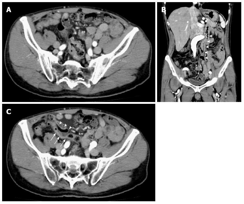
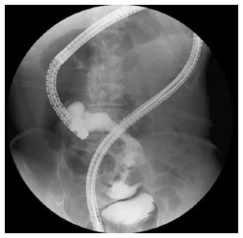
A 60-year-old Japanese man with no significant medical history presented symptoms of melena and dizziness. Because main symptom was dizziness, the patient went to the primary clinic for an examination. Severe anemia was detected using a blood test at the clinic in June 2013. Large intestinal endoscopy was performed as a further examination at the Department of Digestive Medicine in our institute and revealed a mass in the ascending colon. Histological findings of the biopsy specimens indicated colorectal cancer. Preoperative computed tomography (CT) revealed a tumor in the ascending colon with some regional lymph node swelling, but no other organ metastasis was detected. The tumor in the ascending colon was shifted toward the midline of the lower abdomen, placing it in the cavity of the lesser pelvis (Figure 1A). Coronal plane reconstruction of the CT data demonstrated that the descending colon was located at the midline of the abdomen (Figure 1B), and an axial CT image indicated that the sigmoid colon was located under the ascending colon (Figure 1C). A fluoroscopy-assisted endoscopic procedure revealed an abnormal loop of colon in which the sigmoid colon and ascending colon seemed to intersect (Figure 2). Consequently, the preoperative diagnosis was persistent ascending and descending mesocolon.
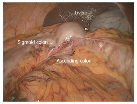
During surgery, we found ascending colon cancer in the pelvis and the ascending colon shifted to the midline without fusion to the retroperitoneum. Notably, the sigmoid colon was shifted to the right abdominal wall, under the ascending colon. An abnormal adhesion between the dorsal aspect of the ascending mesocolon and the ventral aspect of the sigmoid mesocolon was recognized at an early stage of the operation (Figure 3). Accordingly, the descending colon was redundant and included a persistent, long mesocolon. After exfoliating this abnormal adhesion while paying close attention to the prevention of damage to the mesocolon, we performed a laparoscopic right hemicolectomy with radical lymphadenectomy. After resection, a functional end-to-end anastomosis using a linear stapler was safely performed.
The postoperative course was uneventful, and the patient was discharged from the hospital 9 d after the operation. Histopathological examination of the resected specimen demonstrated that the tumor had invaded to the subserosal level and that seven regional lymph nodes were metastatic. Therefore, the patient received adjuvant chemotherapy to reduce the risk of cancer recurrence.
Persistent descending mesocolon results from a failure of the primitive dorsal mesocolon to fuse with the parietal peritoneum during the fifth month of gestation. In the 1960s, this abnormality was reported in the fields of both radiology and gynecology[4,5]. A similar anomaly may appear in the ascending colon after the fifth month of gestation.
In this paper, we report a case involving both ascending and descending mesocolon. To the best of our knowledge, only two case reports of the coexistence of persistent mesocolon of both the ascending and descending colon have been published[6,7]. Kanai et al[6] reported the first case of colonic varices complicated by persistent mesocolon of the ascending and descending colon. Their case was diagnosed using contrast-enhanced irrigoscopy, but the therapy was not described in the report. Ongom et al[7] reported a case of anal protrusion of an ileocolic intussusception with persistent ascending and descending mesocolon, and a laparotomy was performed for this patient. Ours is the first reported case in which laparoscopic colon surgery was performed for persistent ascending and descending mesocolon.
Okada et al[8] published a paper on 13 cases of persistent descending mesocolon in a Japanese institute; this report represents the largest number of cases reported in single institute, to the best of our knowledge. They reported that these 13 cases of abnormalities of sigmoid colon fixation were identified among 543 cases of laparoscopic colectomy for cancer of the descending colon to the rectum. Therefore, the incidence rate of persistent mesocolon with regard to the descending colon was reported to be 2.4% (13/543) in this paper. Moreover, the authors stated that certain anatomical findings should have been noted during the operations, including the fact that the left colon artery, sigmoid colon artery, and superior rectal artery often radially branched from the inferior mesenteric artery and the fact that a marginal vessel ran abnormally due to the unusual mesenteric adhesion.
We usually check not only the axial CT view but also the coronal and sagittal CT views to make an accurate diagnosis before performing laparoscopic colorectal surgery. In this case, the preoperative imaging examination indicated that the descending colon was shifted to the midline, causing the sigmoid colon to be located in the right abdominal cavity and the ascending colon to be located on the sigmoid colon with a mobile cecum. Generally, during laparoscopic right colectomy, the second portion of the duodenum is the most important landmark for the medial approach. In this case, however, the duodenum was not located along the dorsal aspect of the ascending mesocolon, which was anatomically abnormal. Because a medial approach would have been difficult during a surgical resection of the appropriate layers, a lateral approach was selected as the first step. Consequently, a laparoscopic procedure with a high level of safety was able to be performed despite the exceptional anatomical abnormalities.
To the best of our knowledge, no reports have been made on the incidence of the coexistence of both persistent ascending and descending mesocolon. The frequency of the occurrence of persistent ascending mesocolon is expected to be similar to that of persistent descending mesocolon. Therefore, the coexistence of both ascending and descending mesocolon should only be observed rarely in daily clinical practice.
In summary, it is extremely important to consider this anomaly at the time of preoperative CT imaging to ensure a safe operation.
Main symptom of this case was dizziness due to severe anemia by continuous oozing of ascending colon cancer.
Large intestine endoscope examination revealed ascending colon cancer.
Upper gastrointestinal endoscopy was performed to rule out the possibility of stomach bleeding.
Blood test demonstrated low value of hemoglobin and high value of tumor marker.
Because coronal plane reconstruction of the computed tomography (CT) data demonstrated that the descending colon was located at the midline of the abdomen, and an axial CT image indicated that the sigmoid colon was located under the ascending colon, the preoperative diagnosis was persistent ascending and descending mesocolon.
Histopathological examination of the resected specimen demonstrated that the tumor had invaded to the subserosal level and that seven regional lymph nodes were metastatic.
Laparoscopic right hemicolectomy with radical lymphadenectomy was performed and a functional end-to-end anastomosis using a linear stapler was safely performed.
Two important reports in 1960s can help readers better understand the present case (Persistent descending mesocolon. Radiology 1966; 86: 327-331 by Popky et al and Persistent descending mesocolon. Surg Gynecol Obset 1960; 110: 197-2-2 by Morgenstern et al).
Persistent mesocolon is an embryological anomaly that occurs during the final process of intestinal development in organogenesis and it is associated with the absence of fusion between the descending or ascending colon mesentery and the posterior lateral parietal peritoneum, and it appears after 5 mo of gestation.
Although no reports have been made on the incidence of the coexistence of both persistent ascending and descending mesocolon, this clinical pathology always should be remembered at the preoperative CT imaging.
The authors introduce a rare case of the coexistence of both persistent ascending and descending mesocolon. This manuscript shows the distinctive CT image findings of malpositioning of the large intestine.
P- Reviewers: Bini R, O'Dwyer Patrick S- Editor: Ma YJ L- Editor: A E- Editor: Liu XM
| 1. | Ellis H. The gastrointestinal tract. Clinical Anatomy for Students and Junior Doctors. 11th ed. Massecusetts: Blackwell publishing 2006; 91. [Cited in This Article: ] |
| 2. | Balthazar EJ. Congenital positional anomalies of the colon: radiographic diagnosis and clinical implications. II. Abnormalities of fixation. Gastrointest Radiol. 1977;2:49-56. [PubMed] [Cited in This Article: ] |
| 3. | Sadler TW. Langman’s Medical Embryology, 12th ed. In: Taylor C, editor. Philadelphia (PA). 2001; Available from: http://www.doc88.com/p-9049014367116.html. [Cited in This Article: ] |
| 4. | Popky GL, Lapayowker MS. Persistent descending mesocolon. Radiology. 1966;86:327-331. [PubMed] [Cited in This Article: ] |
| 5. | Morgenstern L. Persistent descending mesocolon. Surg Gynecol Obstet. 1960;110:197-202. [PubMed] [Cited in This Article: ] |
| 6. | Kanai M, Tokunaga T, Miyaji T, Mataki N, Okada C, Mitani K, Aono S, Kobari S, Hakozaki Y. Colonic varices as a result of persistent mesocolon of the ascending and descending colon. Endoscopy. 2011;43 Suppl 2:E103-E104. [PubMed] [DOI] [Cited in This Article: ] [Cited by in Crossref: 5] [Cited by in F6Publishing: 9] [Article Influence: 0.7] [Reference Citation Analysis (0)] |
| 7. | Ongom PA, Lukande RL, Jombwe J. Anal protrusion of an ileo-colic intussusception in an adult with persistent ascending and descending mesocolons: a case report. BMC Res Notes. 2013;6:42. [PubMed] [DOI] [Cited in This Article: ] [Cited by in Crossref: 9] [Cited by in F6Publishing: 13] [Article Influence: 1.2] [Reference Citation Analysis (0)] |
| 8. | Okada I, Yamaguchi S, Kondo H, Suwa H, Tashiro J, Ishii T. Laparoscopic colectomy for persistent descending mesocolon: an experience of 13 patients. J Jpn Soc Endosc Surg. 2013;18:459-464. [Cited in This Article: ] |


