Potential roles of the endoplasmic reticulum stress pathway in amyotrophic lateral sclerosis
 Yu-Mi Jeon
 Younghwi Kwon
 Shinrye Lee1
  Hyung-Jun Kim
- 1Dementia Research Group, Korea Brain Research Institute, Daegu, Republic of Korea
- 2Department of Brain and Cognitive Sciences, Daegu Gyeongbuk Institute of Science and Technology (DGIST), Daegu, Republic of Korea
The endoplasmic reticulum (ER) is a major organelle involved in protein quality control and cellular homeostasis. ER stress results from structural and functional dysfunction of the organelle, along with the accumulation of misfolded proteins and changes in calcium homeostasis, it leads to ER stress response pathway such as unfolded protein response (UPR). Neurons are particularly sensitive to the accumulation of misfolded proteins. Thus, the ER stress is involved in neurodegenerative diseases such as Alzheimer’s disease, Parkinson’s disease, prion disease and motor neuron disease (MND). Recently, the complex involvement of ER stress pathways has been demonstrated in experimental models of amyotrophic lateral sclerosis (ALS)/MND using pharmacological and genetic manipulation of the unfolded protein response (UPR), an adaptive response to ER stress. Here, we aim to provide recent evidence demonstrating that the ER stress pathway is an essential pathological mechanism of ALS. In addition, we also provide therapeutic strategies that can help treat diseases by targeting the ER stress pathway.
1. Introduction
Motor neuron disease (MND) refers to a group of rare neurodegenerative diseases that result in a progressive loss of motor function due to selective degeneration of upper motor neurons and/or lower motor neurons (Gagliardi et al., 2021). The cell bodies of motor neurons are in the motor cortex, brainstem or spinal cord, and they control skeletal muscle activity, including breathing, speaking, walking, and swallowing. Motor neuron disease classified two ways: (1) familiar or sporadic, (2) symptomatically. In symptomatically, MND divided into upper motor neuron degeneration (UMN) and lower motor neuron degeneration (LMN; Statland et al., 2015). Motor neuron disease includes amyotrophic lateral sclerosis (ALS), progressive muscular atrophy (DeJesus-Hernandez et al., 2011), progressive lateral sclerosis (PLS), spinal muscular atrophy and Kennedy’s disease (Krivickas, 2003). ALS is the most common and fatal type of MND. The typical feature of ALS is a sudden loss of muscle control and paralysis. Moreover, the average life expectancy of ALS after symptom onset is 2–5 years (van Es et al., 2017; Zhao et al., 2022). More than 90% of ALS cases are sporadic, and 5–10% of ALS patients have a family history of ALS (Hou et al., 2016; Vajda et al., 2017). Sporadic ALS and Familiar ALS also differ in the age of onset. Sporadic ALS cases occurred 58–63, familiar ALS cases are 47–52 approximately (Kiernan et al., 2011). Recent advances in genome sequencing facilitate the identification of new genes associated with ALS, so at least 40 ALS-causing and related genes have been discovered. However, C9orf72, SOD1, TARDBP, and FUS account for half of familial ALS cases (Goutman et al., 2022). There are only 2 FDA-approved medications used to treat ALS, riluzole and edaravone. Riluzole is involved in glutamatergic transmission, and edaravone reduces reactive oxygen species production. The drugs have been shown to prolong life by only a few months or to slow the progression of symptoms, rather than being an actual cure (Jaiswal, 2019; Sadr et al., 2021). The main pathological feature of ALS is abnormal accumulation of misfolded or aggregated proteins in affected neurons and glia. The major roles of the endoplasmic reticulum (ER) are precise folding of newly synthesized proteins and degradation of unfolded or misfolded proteins. Thus, abnormal aggregation of misfolded proteins found in ALS can trigger a stress response called the ER stress pathway. Numerous lines of evidence indicate that ER stress plays an important role in ALS (Ilieva et al., 2007; Vijayalakshmi et al., 2011; Qiu et al., 2013; Atkin et al., 2017; Bugallo et al., 2020).
The ER is an intracellular organelle found in eukaryotic cells. The main functions of the ER are the folding of secreted proteins, participating in calcium signaling as one of the largest calcium stores, and posttranslational modification (Gething and Sambrook, 1992; Helenius et al., 1992; Berridge, 2002). Ca2+ is responsible for the transmission of neuronal depolarization and synaptic activity, and Ca2+ homeostasis and signaling are responsible for proper synaptic plasticity and survival. Thus, disruption of Ca2+ homeostasis affects neurotoxicity and neuronal autophagy and eventually leads to the progression of neurodegenerative disorders (Tong et al., 2018; Mozolewski et al., 2021). The accumulation of unfolded/misfolded proteins within the ER results in stress, which is mitigated by a conserved signaling mechanism known as the highly unfolded protein response (UPR; Moon et al., 2018; Kadowaki and Nishitoh, 2019). However, chronic, or overwhelming ER stress is known to trigger a series of signaling mechanisms that promote cell death and inflammation. Therefore, ER dysfunction and persistent UPR activation may form a central link in the pathogenesis of ALS. In this review, we aim to provide insight into the role and complex interactions of the ER stress pathway and neurodegeneration in ALS.
2. Overview of the endoplasmic reticulum stress pathway
Endoplasmic reticulum is large compartment in eukaryotic cell, and it plays critical role in protein synthesis and folding, Lipid biogenesis, and Ca2+ metabolism (Koch, 1990; Reid and Nicchitta, 2015). These functions are essential for cell. Excessive stress disrupts ER homeostasis, it can lead ER stress. There are several pathways to maintain ER homeostasis: unfolded protein response (UPR), ER-associated degradation (ERAD), and ER-phagy. ERAD recognizes misfolded protein in ER, misfolded proteins are removed by 26S proteasome (Hampton, 2002; Lemus and Goder, 2014; Hwang and Qi, 2018). ER-phagy is one of the autophagy pathways to remove damaged ER (Yang et al., 2021).
The accumulation of unfolded or misfolded proteins in the lumen of the ER induces ER stress, and this stress initiates an interrelated adaptive pathway known as the UPR to restore ER homeostasis. This pathway restores cellular function by halting protein translation, activating signaling pathways that degrade misfolded proteins and increasing the production of molecular chaperones (Mahdi et al., 2016; Nguyen and Uhal, 2016). In addition, activation of the UPR is associated with the generation of proinflammatory conditions (Janssens et al., 2014; Liu et al., 2017). In mammalian cells, the UPR signaling pathway is initiated by three ER membrane-associated sensors: inositol-requiring transmembrane kinase/endoribonuclease 1α (IRE1α), protein kinase RNA-like endoplasmic reticulum kinase (PERK) and activating transcription Factor 6 (ATF6). In normal condition, three sensors interact with 78 kDa glucose-regulated protein/Binding immunoglobulin protein (GRP78/BiP). GRP78/BiP is ER resident chaperon, it acts as a regulator or UPR signaling. Upon misfolded or unfolded protein accumulate in ER, GRP78/BiP dissociated from UPR sensors. After that, PREK and IRE1 form homo-dimerization, respectively, and autophosphorylate (Lee, 2005). Also, ATF6 is moved into Golgi apparatus and cleaved by S1P and S2P proteases. Cleaved ATF6 translocated into nucleus (Figure 1; Guzel et al., 2017).
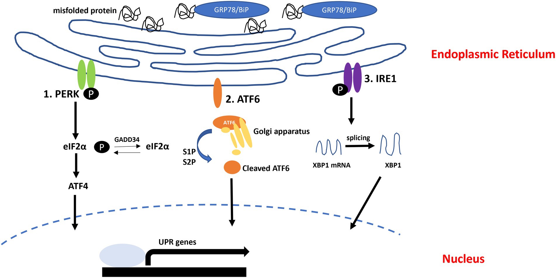
Figure 1. Unfolded protein response (UPR) pathway. In normal condition, GRP78/BiP interact with UPR sensors (PERK, ATF6, and IRE1). When misfolded or unfolded proteins accumulated in ER, GRP78/BiP dissociated from UPR sensors and can bind to misfolded or unfolded proteins. (1) Protein kinase RNA-like endoplasmic reticulum kinase (PERK) autophosphorylates and induces eIF2α phosphorylation. (2) Activating transcription Factor 6 (ATF6) moves into Golgi apparatus and cleaved by S1P and S2P. Cleaved ATF6 moves into nucleus, activates UPR genes. (3) IRE1 autophosphorylates and splices XBP1.
The first branch of the UPR is IRE1, an ER transmembrane sensor. It is perfectly conserved in yeast and animals (Chen and Brandizzi, 2013; Ashford et al., 2021). IRE1 has two isoforms, α and β, and regulates many cellular processes involved in cell survival and apoptosis. Activation of IRE1 is triggered by dissociation of the major ER chaperone, GRP78/BiP, from the luminal domain of IRE1. This dissociation induces dimerization and trans-autophosphorylation, finally triggering the RNase domain activity of IRE1 (Nikawa and Yamashita, 1992; Pincus et al., 2010; Riaz et al., 2020). Eventually, X-box binding protein 1 (XBP1) mRNAs, which are processed by IRE1, translocate to the nucleus and are implicated in the transcriptional upregulation of ER chaperones or adaptive UPR factors for the degradation of misfolded proteins (Yoshida et al., 2001a; Kimata et al., 2004; Junjappa et al., 2018). However, overloaded IRE1 increases the translation of apoptosis-associated proteins by shutting down microRNA (anti-apoptotic) biosynthesis and mediates cell death activation (Upton et al., 2012; Demirel-Yalciner et al., 2021).
Protein kinase RNA-like endoplasmic reticulum kinase is another transducer of unfolded protein response. PERK relieves excessive ER stress by reducing global protein synthesis (Haze et al., 1999; Junjappa et al., 2018; Ohno, 2018). Activation of PERK is triggered by dimerization and autophosphorylation of PERK. Activated PERK shuts off global protein translation by preventing preinitiation complex formation in the ribosome through eukaryotic initiation Factor 2α (eIF2α) phosphorylation at serine 51 (Harding et al., 1999; Humeau et al., 2020). Phosphorylated eIF2α inhibits GDP-GTP exchange, reducing the level of the translationally functional ternary complex and thereby inhibiting translation initiation. However, prolonged attenuation of global protein synthesis leads to disruption of cellular protein homeostasis. Interestingly, PERK-mediated eIF2α phosphorylation has been associated with various diseases, including neurodegenerative disorders. In particular, phosphorylated PERK and phosphorylated eIF2α were significantly increased in postmortem brain tissue of patients with Alzheimer’s disease (AD; Chang et al., 2002; Hoozemans et al., 2005; Lourenco et al., 2015), and similar results were reported in several AD animal models (Devi and Ohno, 2010, 2014). In addition, other neurodegenerative diseases, such as prion disease (Moreno et al., 2012), ALS (Saxena et al., 2009), Parkinson’s disease (Colla et al., 2012) and Huntington’s disease (Leitman et al., 2014), have been associated with the activation of PERK.
The last branch of the UPR is mediated by ATF6. ATF6 is an ER-associated type II membrane-bound protein (Horimoto et al., 2013). When the unfolded proteins accumulate in the ER, ATF6 is translocated from the ER to the Golgi apparatus and is sequentially cleaved by site-specific Site-1 protease and Site-2 protease (Ye et al., 2000). Cleaved ATF6 is translocated to the nucleus to induce transcription of genes encoding ER chaperones and ERAD pathway component genes. In addition, ATF6 also plays a role in the transcriptional upregulation of apoptosis genes (Teske et al., 2011).
Chronic ER stress induces apoptotic cell death via the UPR. Thus, cell death caused by prolonged ER stress is increasingly recognized as a common cause of a wide range of diseases, including neurodegeneration (Moreno et al., 2012; Ismael et al., 2021), inflammation (Bellezza et al., 2014), metabolic disorders (Lemmer et al., 2021), cancer (Samanta et al., 2021), diabetes (Shih et al., 2021), and cardiovascular diseases (Ren et al., 2021). Persistent activation of UPR is associated with ALS and many other neurodegenerative diseases, and recent studies have shown that it is associated with neurons as well as non-neuronal cells such as astrocytes and microglia (Figure 2; Ito et al., 2009; Walker and Atkin, 2011; Sims et al., 2022). Therefore, inhibition of the UPR may be a promising therapeutic strategy for these diseases.
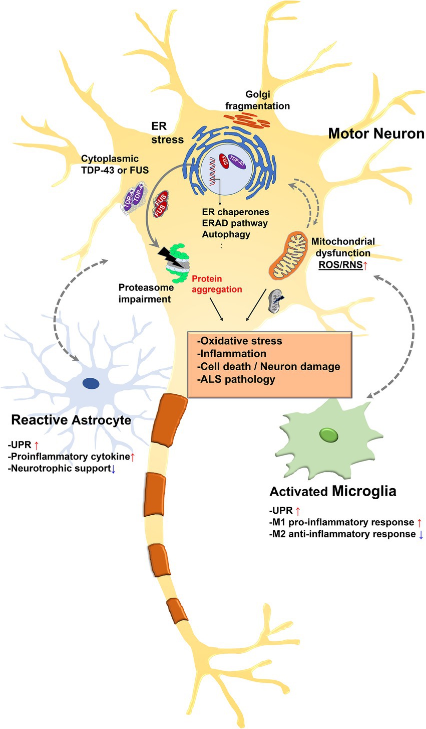
Figure 2. Relationship between endoplasmic reticulum (ER) stress and amyotrophic lateral sclerosis (ALS)-associated pathology. Misfolded proteins accumulate in the ER lumen and this accumulation contributes to ER stress. ER stress triggers the UPR to restore protein homeostasis. However, chronic stress leads to a cascade of intracellular death and inflammatory signal. Some nuclear ALS disease proteins such as TAR DNA-binding protein 43 (TDP-43) or fused in sarcoma (FUS) are depleted in the nucleus and accumulate in the cytoplasm in pathologic condition. Pathologic TDP-43 or FUS are existed as hyperphosphorylated and ubiquitinated form. Persistent activation of the stress response leads to ER dysfunction and ubiquitin proteasome system (UPS) impairment, leading to reactive oxygen species (ROS) and RNS release in mitochondria. This eventually leads to insoluble cytoplasmic aggregation and impairment of mitochondrial and ER function. Furthermore, UPR-related genes are upregulated in astrocytes and microglia along with inflammatory genes. Specifically, UPR activation reduces the ability of glia to support synapse and is associated with neuronal cell death. In addition, glia could transmit ER stress to neurons, which in turn may exacerbate ER stress mediated neuronal damage.
3. The relationship between ER stress and ALS-associated genes
3.1. Superoxide dismutase 1
Superoxide dismutase 1 (SOD1) is an antioxidant enzyme that protects against oxidative stress in eukaryotic cells (Trist et al., 2021). An association between SOD1 and familial ALS (fALS) was identified in 1993 (Rosen et al., 1993). ALS patients associated with SOD1 mutation contain SOD1-immunoreactive inclusions (Bruijn et al., 1998). In fALS cases, up to 20% of cases have SOD1 mutations; to date, more than 180 mutations have been identified in ALS cases (Hayashi et al., 2016). SOD1G93A mice were the first transgenic mice used as an ALS model, and their phenotype is very similar to that of ALS in humans (Gurney et al., 1994). The ALS-associated SOD1 mutation induces several pathological features, such as oxidative stress (Ferrante et al., 1997), mitochondrial dysfunction (Mattiazzi et al., 2002; Damiano et al., 2006), and prion-like propagation of disease proteins (Grad et al., 2014). Furthermore, ER stress is an important SOD1-related pathology (Nagata et al., 2007; Nishitoh et al., 2008). In SOD1G93A mutant mice, increased phosphorylation of PERK and eIF2α was observed. Increased ER stress markers were found at the mid-to-late presymptomatic stage in SOD1G93A transgenic mice. Following the response of PERK/eIF2α phosphorylation, Caspase12 expression is also increased. Caspase 12 is located on the outer surface of the ER, and its activation elicits the proapoptotic caspase cascade. Thus, these results indicate that ER stress leads to cell death in mutant SOD1 mice (Nagata et al., 2007).
Endoplasmic reticulum-associated degradation (ERAD) targets misfolded proteins in ER, misfolded proteins transported from ER to cytosol, where they are degraded by ubiquitin proteasome system (UPS) under activated UPR conditions. Inhibition of ERAD disrupts the cytosolic transport of misfolded proteins, and they accumulate in the ER and induce ER stress (Kopito, 1997; Tsai et al., 2002). Moreover, mutant SOD1 directly binds to Derlin-1 in the ER (Nishitoh et al., 2008) and that Derlin-1 is an important protein in ERAD-mediated translocation from the ER to the cytosol (Lilley and Ploegh, 2004; Ye et al., 2004). The interaction between the SOD1 mutant and Derlin-1 inhibits the translocation of misfolded proteins from the ER to the cytosol, and misfolded proteins accumulate in the ER. The mutant SOD1-Derlin1 interaction induces IRE1/ASK1 signaling and cell death (Nishitoh et al., 2008).
It is known that phosphorylation of PERK induces ATF4 expression through eIF2α phosphorylation. Consistently, SOD1 mutant ALS mice showed upregulated ATF4 expression compared to control mice (Kikuchi et al., 2006). Moreover, upregulated UPR genes were related to accumulation of SOD1 in the ER. Under normal conditions, BiP interacts with UPR sensing proteins such as PERK, ATF6, and IRE-1. In SOD1 mutant ALS mice, the binding affinity between BiP and other UPR sensors is increased (Kikuchi et al., 2006).
Protein disulfide isomerase A1 (PDIA1, refer to PDI) is a chaperone that localizes to the ER. ER stress upregulates PDI, and it plays a protective role in protein misfolding and aggregation (Parakh and Atkin, 2015). PDI interacts with misfolded proteins rather than native proteins in the ER through hydrophobic interactions (Parakh and Atkin, 2015). PDI promotes the translocation of misfolded proteins from the ER to the cytosol via ERAD, and misfolded proteins are degraded by the UPS (Molinari et al., 2002; Lee et al., 2010). Overexpression of PDI reduces mutant SOD1 aggregation in NSC-34 motor neuron like cell lines. Moreover, the expression of BiP, CHOP, and PERK phosphorylation was decreased by PDI overexpression in NSC-34 cells. Several studies have reported that ER stress in the SOD1 ALS model induces apoptotic cell death. PDI overexpression mitigates SOD1 mutant-induced cell death, and knockdown of PDI increased mutant SOD1 inclusions in NSC-34 cells (Walker et al., 2010). Based on this, ER stress-related chaperones might be mitigate mutant SOD1-induced ER stress.
3.2. Transactive response DNA-binding protein 43
Cytoplasmic aggregation of TAR DNA-binding protein 43 (TDP-43) is related to many neurodegenerative diseases referred to as TDP-43 proteinopathies (Kwong et al., 2007; Scotter et al., 2015). Cytoplasmic TDP-43 aggregate is a major pathologic hallmark of ALS (Neumann et al., 2006). More than 97% of ALS patients have TDP-43-positive inclusions in their affected neurons and glia (Mackenzie et al., 2007; Maekawa et al., 2009). Several studies have shown that overexpression of wild-type or ALS-associated mutant TDP-43 causes neurotoxicity (Ash et al., 2010; Kabashi et al., 2010; Li et al., 2010; Wils et al., 2010; Cascella et al., 2016). Immunoelectron microscopy data showed that TDP-43 was decreased in the nucleus and increased in the ER in ALS patients compared with controls (Sasaki et al., 2010). This research indicated that nuclear depletion of TDP-43 was related to TDP-43 accumulation in the ER (Sasaki et al., 2010; de Mena et al., 2021). Furthermore, overexpression of disease-associated TDP-43 mutations (D169G, G294A, A315T, Q331K, M337V, and N390D) increased cytoplasmic accumulation and induced UPR more than wild-type TDP-43 overexpression (Walker et al., 2013; Wang et al., 2015). Overexpression of TDP-43 also upregulates CHOP mRNA levels, and TDP-43-induced cell death is mediated by CHOP (Suzuki and Matsuoka, 2012). CHOP expression is regulated by the UPR such as PERK/eIF2α/ATF4 pathway. A previous study showed that TDP-43-induced cell death is also related to Bim activation (Suzuki et al., 2011) and that CHOP acts as a transcription factor for the Bim expression (Puthalakath et al., 2007). Moreover, CHOP expression is more strongly induced by mutant TDP-43 compared to wild type TDP-43 (Walker et al., 2013). These results suggest that TDP-43 accumulation in the ER can lead to ER stress and ER stress is implicated in TDP-43 induced neurotoxicity.
Many studies have shown that wildtype or mutant TDP-43 can increase eIF2α phosphorylation. Overexpression of TDP-43 in Drosophila nervous system increased the phosphorylation of eIF2α (Ser51; Kim et al., 2014). Moreover, increased eIF2α phosphorylation is essential in TDP-43-mediated neurotoxicity in Drosophila. Inhibition of PERK using GSK2606414 (PERK inhibitor) reduced eIF2α phosphorylation and rescued TDP-43-mediated neurotoxicity in Drosophila and mammalian neurons. Inhibition of GADD34 (eIF2α phosphatase) also enhances eIF2α phosphorylation and accelerates TDP-43-induced neurotoxicity (Kim et al., 2014). In addition, overexpression of TDP-43 in SH-SY5Y human neuroblastoma cells increased eIF2α phosphorylation, and increased eIF2α phosphorylation upregulates the transcription levels of ER stress-related genes such as ATF4, CHOP, and GADD34. Similarly, a reduction in eIF2α phosphorylation restores the transcription level of ER stress-related genes and mitigates TDP-43-induced cell death in SH-SY5Y cells (Jeon et al., 2021).
Protein kinase R (PKR or eukaryotic translation initiation factor-2 alpha kinase 2, EIF2AK2) regulates diverse cellular responses and is also associated with ER stress-related apoptosis through eIF2α phosphorylation, CHOP, and the ATF4 pathway. Under ER stress conditions, activation of PKR directly phosphorylates eIF2α in a PERK-independent manner and induces CHOP and ATF4 expression (Lee et al., 2007). Previous study indicated that knockdown of TDP-43 in astrocytes induces a robust increase in eIF2α phosphorylation, which is attenuated by PKR inhibition (LaRocca et al., 2019). Furthermore, TDP-43A315T and TDP-43Q331K promote neuronal toxicity in SH-SY5Y cells via ER stress pathway, such as eIF2α phosphorylation, CHOP, and GRP78 (Wang et al., 2015; Hu et al., 2019). In ALS patients who have the TDP-43A315T mutation, the ER stress-related chaperone GRP78 is increased compared to that of controls (Wang et al., 2015).
Activation of the IRE-1 mitigates ER stress mediated cytotoxicity via upregulation of XBP1 splicing (Yoshida et al., 2001a; Dafinca et al., 2021). TDP-43M337V expression in rat neurons decreased spliced XBP1. Moreover, ubiquitin aggregation and Golgi fragmentation were also observed in the neurons of TDP-43M337V transgenic rats (Tong et al., 2012). On the other hand, another ALS-related mutant (TDP-43Q331K, TDP-43A315T) overexpression increased XBP1 and ATF6 expression compared with wild-type TDP-43 overexpression in N2a neuronal cells (Walker et al., 2013). Although relationship between XBP1 and TDP-43 mutant is controversial, TDP-43-related ER stress is associated with IRE1/XBP1 signaling pathways.
3.3. Fused in sarcoma
Fused in sarcoma (Li et al., 2010) was initially identified as a transcriptional regulator in human malignant liposarcomas in 1993 (Rabbitts et al., 1993). In 2009, a FUS mutation was identified in ALS patients (Kwiatkowski et al., 2009; Vance et al., 2009). In ALS patients, ~ 4% of FUS mutations are found in fALS, ~ 1% of mutations are found in sporadic ALS cases, and to date, more than 50 FUS mutations have been shown in ALS patients (Zou et al., 2017; Kim et al., 2020). FUS aggregation were observed in the spinal cord of ALS patients with FUS mutation, and FUS inclusion was colocalized with TDP-43 and ubiquitin (Deng et al., 2010). FUS is a DNA/RNA-binding protein; similar to TDP-43. FUS predominantly localizes to the nucleus (Lattante et al., 2013) and can shuttle between the nucleus and cytoplasm (Zinszner et al., 1997). Disease associated FUS mutations facilitate the cytoplasmic mislocalization of FUS. FUSP525L and FUSR522G mutants impair transportin mediated nuclear transport of FUS (Dormann et al., 2010).
Amyotrophic lateral sclerosis (ALS) linked mutant FUS induce ER stress, and overexpression of mutant FUS colocalized with calreticulin (as a marker of ER) in NSC-34 motor neuron cells. Overexpression of mutant FUS increases the overlap with calreticulin compared to wildtype FUS overexpression, and cytoplasmic mislocalization of FUS is also increased compare with wildtype FUS in NSC-34 (Farg et al., 2012). Moreover, mutant FUS expression induces XBP1 splicing and CHOP expression (Farg et al., 2012). These results indicated that cytoplasmic aggregation of FUS correlates with ER stress.
Fused in sarcoma (FUS) is also related to PDI. Overexpression of the FUS mutant in NSC-34 cells increased PDI expression compared to wildtype FUS overexpression. ALS patients who have the FUSR521C mutation showed colocalization of FUS with PDI in the spinal cord (Farg et al., 2012). A recent study showed that overexpression of PDI reduces mutant FUS-induced ER stress. Overexpression of mutant FUS induces nuclear translocation of CHOP. However, overexpression of PDI decreases the nuclear immunoreactivity of CHOP in mutant FUS-overexpressing cells (Parakh et al., 2021). FUS is a RNA-binding protein with a prion-like domain that can form aggregates (Gitler and Shorter, 2011). A previous study showed that PDI can interact with prion-like proteins, suggesting that PDI can bind FUS through prion-like domains (Watts et al., 2009). Thus, enhancing the interaction between FUS and PDI might be the promising therapeutic strategy for FUS mediated neurodegeneration.
3.4. Chromosome 9 open reading frame 72
The expansion of the GGGGCC (G4C2) hexanucleotide in the chromosome 9 open reading frame 72 (C9orf72) gene accounts for 10% of ALS patients. Among them, ~ 40% are fALS cases (DeJesus-Hernandez et al., 2011; Yang et al., 2020), and 5 ~ 10% are sporadic cases (Umoh et al., 2016). There are several pathological features in C9orf72-related diseases, including toxic gain or loss of function in C9orf72, dipeptide repeat (DPR) proteins generated by repeat-associated non-ATG (RAN) translation, and transcriptional silencing (Dafinca et al., 2021). It is known that RAN translation of the C9orf72 gene produces five DPRs, glycine–alanine (GA), glycine-proline (Kim et al., 2022), glycine-arginine (GR), proline-alanine (PA), and proline-arginine (PR; Freibaum and Taylor, 2017).
Transcriptome analysis in the cerebellum and frontal cortex of C9orf72-ALS patients revealed altered UPR-related gene expression such as ATF4 and CHOP (Freibaum and Taylor, 2017). Phosphorylation of PERK, another marker for ER stress, was also increased in the brain of C9orf72-ALS patient. Expression of poly-GA proteins induces neuronal toxicity through ER stress in mouse primary cortical neurons (Zhang et al., 2014). Overexpression of poly-PR using lentivirus in primary cortical neurons showed that ER stress-related genes were highly upregulated by PR, and PR expressing cells also showed increased neuronal toxicity. Moreover, the mRNA levels of ATF4, CHOP, and GADD34 were also upregulated by poly-PR expression in K562 cell lines (Kramer et al., 2018). Consistent with this research, treatment of SH-SY5Y cells and primary cortical neurons with the poly-PR peptide increased the neuronal toxicity and transcription levels of ATF4 and CHOP in a dose-dependent manner (Wang et al., 2019). Interestingly, recent research has revealed a relationship between RAN translation of C9orf72 and eIF2α phosphorylation (Cheng et al., 2018). Repeat-associated non-ATG (RAN) translation of C9ORF72 hexanucleotide repeats are increased by excessive phosphorylation of eIF2α. All these data indicated that ER stress is related to C9ORF72-mediated neurodegeneration.
3.5. Others
3.5.1. Optineurin
Optineurin (OPTN) is associated with several cellular functions, such as the inflammatory response, autophagy, and Golgi maintenance (Toth and Atkin, 2018). More than 20 mutations in OPTN are associated with ALS. Unlike other ALS-causing mutation, TDP-43, SOD1, and FUS, OPTN mutations do not facilitate protein aggregation (Markovinovic et al., 2017). Overexpression of the ALS-associated OPTNE478G mutation increased the annexin V/7-AAD-positive cells in NSC-34, indicating that mutant OPTN induces cell death (Sayyad et al., 2017). A 2-base pair insertion in OPTN (619-692insAG or 2 bpIsn-OPTN) is associated with both ALS and glaucoma. 2 bpIsn-OPTN increased caspase-3 activation and ER stress in NSC-34 cells. Overexpression of 2 bpIsn-OPTN in NSC-34 cells upregulated the transcription of CHOP and XBP1. Moreover, treatment with an ER stress inhibitor decreased ER stress-related gene expression and reduced cell death. 2 bpIsn-OPTN is predominantly localized in the nucleus, in contrast to wild-type OPTN mainly localized in the cytoplasm (Medchalmi et al., 2021). Loss of OPTN in mouse and MEF cells also induced upregulation of ER stress pathway (Ramachandran et al., 2021). OPTN knockout cells are more susceptible to cytotoxicity of ER stress inducers (tunicamycin and thapsigargin), and UPR-related genes were also highly upregulated. Moreover, basal levels of PERK and IRE1 were significantly higher in OPTN knockout MEFs. OPTN deletion also increased the mRNA level of CHOP in the tunicamycin treated mouse brain (Ramachandran et al., 2021). These results suggest that loss of OPTN can induce cell death through ER stress pathway.
3.5.2. Ubiquitin-like protein ubiquilin2
The ubiquitin-like protein ubiquilin2 (UBQLN2) mutation is related to fALS cases, but the pathological features of UBQLN2 are largely unknown. However, UBQNL2 is involved in ERAD mediated protein degradation via UPS. Moreover, the best described UBQLN2-associated pathologies are the UPS and Autophagy impairment (Renaud et al., 2019). The viral expression of ALS linked UBQLN2 mutations (P497H, P497S, and P506T) in mouse brain caused reduction of motor capacity and accumulation of UBQLN2 containing inclusions (Ceballos-Diaz et al., 2015). Moreover, UBQLN2P497H and UBQLN2P506T overexpression in Neuro2A cells upregulates ER stress mediators such as XBP1 and CHOP compared to wildtype UBQLN2 (Halloran et al., 2020). Taken together, these data indicate that ER stress might be implicated in UBQLN2 induced neurodegeneration.
4. Endoplasmic reticulum stress as a therapeutic target for ALS
4.1. Salubrinal
Salubrinal is selective inhibitor of eIF2α phosphatase (Lu et al., 2022). Salubrinal upregulates eIF2α phosphorylation and reduces ER stress-induced cytotoxicity in PC12 cells (Boyce et al., 2005). In SOD1G93A- and SOD1G85R-transfected N2a cells, salubrinal treatment prevented SOD1-induced cell death through regulation of eIF2α phosphorylation (Oh et al., 2008). Furthermore, salubrinal treatment reduced TDP-43-induced neurotoxicity in C. elegans and zebrafish. Mutant TDP-43-expressing C. elegans and zebrafish showed motor neuron defects, and salubrinal treatment attenuated TDP-43 induced motor neuron degeneration (Vaccaro et al., 2013). Salubrinal-treated SOD1G93A mice showed alleviation of disease progression, such as increased survival ability and reinnervation of neuromuscular junctions (NMJs; Saxena et al., 2009).
4.2. Arimoclomol
Arimoclomol, called BRX-220, is a co-inducer of heat-shock protein (Hargitai et al., 2003) and has been shown to be a therapeutic candidate in several diseases (Fog et al., 2018). Several studies have suggested that treatment with arimoclomol in ALS models prevents disease-associated pathology. Arimoclomol-treated SOD1G93A mice had an increased lifespan (Kieran et al., 2004), and the delayed overall disease progression (Kalmar et al., 2008). The heat shock response (HSR) is essential for recovery of proteotoxic damage and associated with refolding and degradation of misfolded or unfolded proteins (Westerheide and Morimoto, 2005). Arimoclomol treatment restored HSP70 expression which is decreased by mutant SOD1 and prevented ER stress-induced motor neuron death in SOD1G93A mice (Kieran et al., 2004; Kalmar et al., 2008). However, phase III clinical trial for arimoclomol against ALS failed in 2021. The ORARIALS-01 phase 3 trial (Clinicaltrialsgov identifier: NCT03491462) investigated 245 adults with ALS across North America and Europe receiving arimoclomol or placebo once daily for 76 weeks. As a result of evaluating the effect of arimoclomol through an integrated evaluation of function and survival, it was announced that the goal was not achieved1.
4.3. PRE-084
PRE-084 is a sigma-1 receptor agonist. The sigma-1 receptor is a transmembrane receptor that localizes to the ER and mitochondria-associated ER membrane (MEM; Mancuso and Navarro, 2017). It has neuroprotective effects that regulate Ca2+ signaling, mitochondrial function, and ER stress (Hayashi and Su, 2007; Sharma et al., 2021). The sigma-1 receptor can translocate between the ER and MAM junction and plays a critical role in ER-mitochondria communication. The sigma-1 receptor directly binds to Bip, and it respond to ER stress and regulate Ca2+ homeostasis (Hayashi and Su, 2007). Several studies have shown that sigma-1 receptor is associated with ALS pathology. In SOD1G93A mice, deletion of the sigma-1 receptor accelerated motor neuron dysfunction and decreased longevity (Mavlyutov et al., 2013). Another study showed that loss of the sigma-1 receptor affected the mitochondria-ER connection, leading to ER stress and dysfunction of Ca2+ signaling in culture mouse motor neurons (Bernard-Marissal et al., 2015). Furthermore, mutation of the sigma-1 receptor was found in ALS patients, and this mutation induced neuronal cell death through ER stress in NSC-34 motor neuron-like cells (Al-Saif et al., 2011; Bernard-Marissal et al., 2015). PRE-084 treatment in SOD1G93A mice rescued motor neuron dysfunction, including survival, through PCK activation (Mancuso et al., 2012). SA4503, another sigma-1 receptor agonist, also reduced SOD1G93A-induced cell death and extended the survival time of SOD1G93A mice (Ono et al., 2014).
4.4. Sephin1
Regulators of the PERK pathway (Salubrinal, Sephin1 and Guanabenz) inhibit the protein phosphatase complex to prevent dephosphorylation of eIF2α, a negative feedback loop that regulates protein translation during ER stress. Selphin1 is a selective inhibitor of GADD34 (PPP1R15A), a stress-induced regulatory subunit of the protein phosphatase 1 complex that dephosphorylates eIF2α (Das et al., 2015; Chen et al., 2021). Sephin1 has shown a strong effect in preventing motor neuron degeneration in in vitro and in vivo ALS models (Das et al., 2015; Chen et al., 2019). Sephin1 prevented the accumulation of the insoluble SOD1 and decreased the ER stress markers including Bip, CHOP, and Xbp-1 in spinal cords of SOD1G93A mice. Thus, Sephin1 mitigated motor deficits, motor neuron loss, and molecular defects in SOD1 mutant mice (Das et al., 2015).
4.5. Guanabenz
Guanabenz is an FDA-approved alpha-2 adrenergic receptor agonist used as an antihypertensive agent. Guanabenz has been shown to prevent the accumulation of misfolded proteins and ER overload by regulating protein synthesis (Tsaytler et al., 2011). An in vitro study showed that Guanabenz prolonged eIF2α phosphorylation and modulated the rate of protein production in stressed human cells. This provided strong evidence that restoring protein homeostasis in stressed cells could be a drug target for ALS (Tsaytler et al., 2011). According to an in vivo study using the SOD1 mutant transgenic mice, Guanabenz attenuated ER stress due to prolonged eIF2α phosphorylation or downregulated the expression of proapoptotic proteins, thereby delaying disease onset, extending lifespan, and reducing motor neuron loss in mice (Jiang et al., 2014; Wang et al., 2014). Moreover, a phase II randomized clinical trial in ALS patients showed that a combination of guanabenz with riluzole slowed disease progression in early ALS patients with bulbar onset (Dalla Bella et al., 2021). As described above, many drugs that directly target UPR are summarized in a table (Table 1).
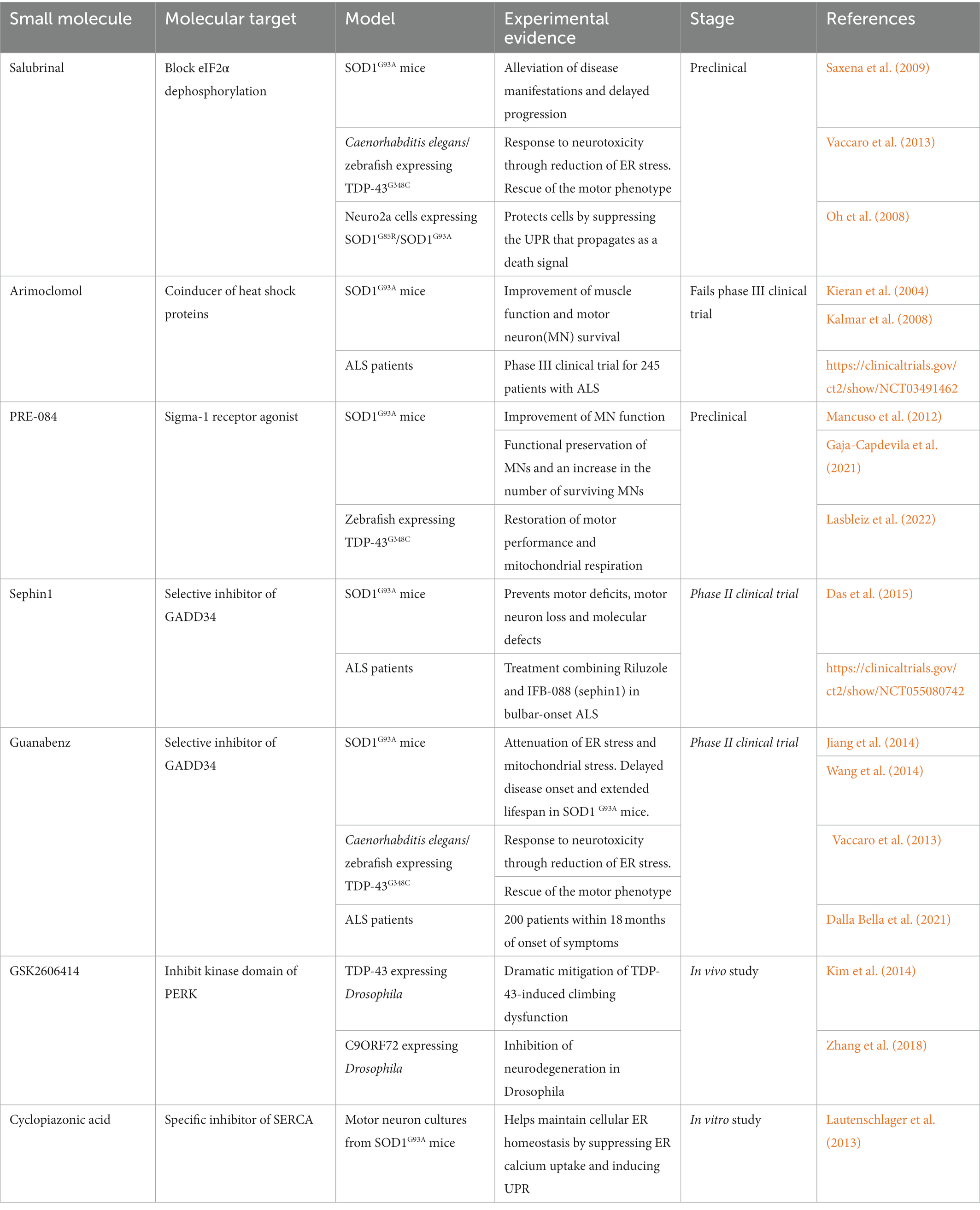
Table 1. Small molecules targeting the unfolded protein response (UPR) pathway for amyotrophic lateral sclerosis (ALS) treatment.
5. Conclusion
Growing evidence indicates that the ER stress pathway is strongly implicated in the pathogenesis of ALS. Although the UPR is a cellular adaptive and protective response to overwhelming ER stress, sustained stress can result in an imbalance in protein homeostasis, resulting in increased ROS production. The UPR pathway is facilitated by the IRE1, PERK and ATF6 branches, which are important for maintaining protein homeostasis, and this protective role is becoming more important in ALS/MND. It is important to note that the UPR pathway may be affected equally or differently for each cell type involved. For example, microglia adopt an M2-like neuroprotective state early in disease but are likely to transition to an M1-like toxic state as ALS progresses (Liao et al., 2012; Chiu et al., 2013; Clarke and Patani, 2020).Therefore, a clearer and improved understanding of the relevant mechanisms of genetic and protein homeostatic dysfunction associated with the more complex ALS/MND will be essential and important for developing small molecule therapeutics that effectively target the UPR mediated ALS pathogenesis.
Author contributions
Y-MJ, YK, and SL provided ideas for the project and participated in data collection. Y-MJ, YK, and H-JK wrote the paper. All authors contributed to the article and approved the submitted version.
Funding
This work was supported by the KBRI Research Program of the Ministry of Science, ICT and Future Planning (22-BR-02-04; 22-BR-03-02) and the National Research Foundation of Korea (NRF) grant funded by the Korean government (MSIT; 2020R1A2C4002366; 2021R1C1C1008688).
Conflict of interest
The authors declare that the research was conducted in the absence of any commercial or financial relationships that could be construed as a potential conflict of interest.
Publisher’s note
All claims expressed in this article are solely those of the authors and do not necessarily represent those of their affiliated organizations, or those of the publisher, the editors and the reviewers. Any product that may be evaluated in this article, or claim that may be made by its manufacturer, is not guaranteed or endorsed by the publisher.
Footnotes
References
Al-Saif, A., Al-Mohanna, F., and Bohlega, S. (2011). A mutation in sigma-1 receptor causes juvenile amyotrophic lateral sclerosis. Ann. Neurol. 70, 913–919. doi: 10.1002/ana.22534
Ash, P. E., Zhang, Y. J., Roberts, C. M., Saldi, T., Hutter, H., Buratti, E., et al. (2010). Neurotoxic effects of TDP-43 overexpression in C. elegans. Hum. Mol. Genet. 19, 3206–3218. doi: 10.1093/hmg/ddq230
Ashford, B. A., Boche, D., Cooper-Knock, J., Heath, P. R., Simpson, J. E., and Highley, J. R. (2021). Review: microglia in motor neuron disease. Neuropathol. Appl. Neurobiol. 47, 179–197. doi: 10.1111/nan.12640
Atkin, J. D., Farg, M. A., Turner, B. J., Tomas, D., Lysaght, J. A., Nunan, J., et al. (2017). Withdrawal: induction of the unfolded protein response in familial amyotrophic lateral sclerosis and association of protein-disulfide isomerase with superoxide dismutase 1. J. Biol. Chem. 292:12007. doi: 10.1074/jbc.A117.603393
Bellezza, I., Grottelli, S., Mierla, A. L., Cacciatore, I., Fornasari, E., Roscini, L., et al. (2014). Neuroinflammation and endoplasmic reticulum stress are coregulated by cyclo(his-pro) to prevent LPS neurotoxicity. Int. J. Biochem. Cell Biol. 51, 159–169. doi: 10.1016/j.biocel.2014.03.023
Bernard-Marissal, N., Medard, J. J., Azzedine, H., and Chrast, R. (2015). Dysfunction in endoplasmic reticulum-mitochondria crosstalk underlies SIGMAR1 loss of function mediated motor neuron degeneration. Brain 138, 875–890. doi: 10.1093/brain/awv008
Berridge, M. J. (2002). The endoplasmic reticulum: a multifunctional signaling organelle. Cell Calcium 32, 235–249. doi: 10.1016/S0143416002001823
Boyce, M., Bryant, K. F., Jousse, C., Long, K., Harding, H. P., Scheuner, D., et al. (2005). A selective inhibitor of eIF2alpha dephosphorylation protects cells from ER stress. Science 307, 935–939. doi: 10.1126/science.1101902
Bruijn, L. I., Houseweart, M. K., Kato, S., Anderson, K. L., Anderson, S. D., Ohama, E., et al. (1998). Aggregation and motor neuron toxicity of an ALS-linked SOD1 mutant independent from wild-type SOD1. Science 281, 1851–1854. doi: 10.1126/science.281.5384.1851
Bugallo, R., Marlin, E., Baltanás, A., Toledo, E., Ferrero, R., Vinueza-Gavilanes, R., et al. (2020). Fine tuning of the unfolded protein response by ISRIB improves neuronal survival in a model of amyotrophic lateral sclerosis. Cell Death Dis. 11:397. doi: 10.1038/s41419-020-2601-2
Cascella, R., Capitini, C., Fani, G., Dobson, C. M., Cecchi, C., and Chiti, F. (2016). Quantification of the relative contributions of loss-of-function and gain-of-function mechanisms in TAR DNA-binding protein 43 (TDP-43) Proteinopathies. J. Biol. Chem. 291, 19437–19448. doi: 10.1074/jbc.M116.737726
Ceballos-Diaz, C., Rosario, A. M., Park, H. J., Chakrabarty, P., Sacino, A., Cruz, P. E., et al. (2015). Viral expression of ALS-linked ubiquilin-2 mutants causes inclusion pathology and behavioral deficits in mice. Mol. Neurodegener. 10:25. doi: 10.1186/s13024-015-0026-7
Chang, R. C., Wong, A. K., Ng, H. K., and Hugon, J. (2002). Phosphorylation of eukaryotic initiation factor-2alpha (eIF2alpha) is associated with neuronal degeneration in Alzheimer's disease. Neuroreport 13, 2429–2432. doi: 10.1097/00001756-200212200-00011
Chen, Y., and Brandizzi, F. (2013). IRE1: ER stress sensor and cell fate executor. Trends Cell Biol. 23, 547–555. doi: 10.1016/j.tcb.2013.06.005
Chen, Y., Kunjamma, R. B., Weiner, M., Chan, J. R., and Popko, B. (2021). Prolonging the integrated stress response enhances CNS remyelination in an inflammatory environment. elife 10:e65469. doi: 10.7554/eLife.65469
Chen, Y., Podojil, J. R., Kunjamma, R. B., Jones, J., Weiner, M., Lin, W., et al. (2019). Sephin1, which prolongs the integrated stress response, is a promising therapeutic for multiple sclerosis. Brain 142, 344–361. doi: 10.1093/brain/awy322
Cheng, W., Wang, S., Mestre, A. A., Fu, C., Makarem, A., Xian, F., et al. (2018). C9ORF72 GGGGCC repeat-associated non-AUG translation is upregulated by stress through eIF2alpha phosphorylation. Nat. Commun. 9:51. doi: 10.1038/s41467-017-02495-z
Chiu, I. M., Morimoto, E. T., Goodarzi, H., Liao, J. T., O'Keeffe, S., Phatnani, H. P., et al. (2013). A neurodegeneration-specific gene-expression signature of acutely isolated microglia from an amyotrophic lateral sclerosis mouse model. Cell Rep. 4, 385–401. doi: 10.1016/j.celrep.2013.06.018
Clarke, B. E., and Patani, R. (2020). The microglial component of amyotrophic lateral sclerosis. Brain 143, 3526–3539. doi: 10.1093/brain/awaa309
Colla, E., Jensen, P. H., Pletnikova, O., Troncoso, J. C., Glabe, C., and Lee, M. K. (2012). Accumulation of toxic α-synuclein oligomer within endoplasmic reticulum occurs in α-synucleinopathy in vivo. J. Neurosci. 32, 3301–3305. doi: 10.1523/JNEUROSCI.5368-11.2012
Dafinca, R., Barbagallo, P., and Talbot, K. (2021). The role of mitochondrial dysfunction and ER stress in TDP-43 and C9ORF72 ALS. Front. Cell. Neurosci. 15:653688. doi: 10.3389/fncel.2021.653688
Dalla Bella, E., Bersano, E., Antonini, G., Borghero, G., Capasso, M., Caponnetto, C., et al. (2021). The unfolded protein response in amyotrophic later sclerosis: results of a phase 2 trial. Brain 144, 2635–2647. doi: 10.1093/brain/awab167
Damiano, M., Starkov, A. A., Petri, S., Kipiani, K., Kiaei, M., Mattiazzi, M., et al. (2006). Neural mitochondrial Ca2+ capacity impairment precedes the onset of motor symptoms in G93A cu/Zn-superoxide dismutase mutant mice. J. Neurochem. 96, 1349–1361. doi: 10.1111/j.1471-4159.2006.03619.x
Das, I., Krzyzosiak, A., Schneider, K., Wrabetz, L., D'Antonio, M., Barry, N., et al. (2015). Preventing proteostasis diseases by selective inhibition of a phosphatase regulatory subunit. Science 348, 239–242. doi: 10.1126/science.aaa4484
de Mena, L., Lopez-Scarim, J., and Rincon-Limas, D. E. (2021). TDP-43 and ER stress in neurodegeneration: friends or foes? Front. Mol. Neurosci. 14:772226. doi: 10.3389/fnmol.2021.772226
DeJesus-Hernandez, M., Mackenzie, I. R., Boeve, B. F., Boxer, A. L., Baker, M., Rutherford, N. J., et al. (2011). Expanded GGGGCC hexanucleotide repeat in noncoding region of C9ORF72 causes chromosome 9p-linked FTD and ALS. Neuron 72, 245–256. doi: 10.1016/j.neuron.2011.09.011
Demirel-Yalciner, T., Sozen, E., and Ozer, N. K. (2021). Endoplasmic reticulum stress and miRNA impairment in aging and age-related diseases. Front. Aging 2:790702. doi: 10.3389/fragi.2021.790702
Deng, H. X., Zhai, H., Bigio, E. H., Yan, J., Fecto, F., Ajroud, K., et al. (2010). FUS-immunoreactive inclusions are a common feature in sporadic and non-SOD1 familial amyotrophic lateral sclerosis. Ann. Neurol. 67, 739–748. doi: 10.1002/ana.22051
Devi, L., and Ohno, M. (2010). Phospho-eIF2α level is important for determining abilities of BACE1 reduction to rescue cholinergic neurodegeneration and memory defects in 5XFAD mice. PLoS One 5:e12974. doi: 10.1371/journal.pone.0012974
Devi, L., and Ohno, M. (2014). PERK mediates eIF2α phosphorylation responsible for BACE1 elevation, CREB dysfunction and neurodegeneration in a mouse model of Alzheimer's disease. Neurobiol. Aging 35, 2272–2281. doi: 10.1016/j.neurobiolaging.2014.04.031
Dormann, D., Rodde, R., Edbauer, D., Bentmann, E., Fischer, I., Hruscha, A., et al. (2010). ALS-associated fused in sarcoma (FUS) mutations disrupt Transportin-mediated nuclear import. EMBO J. 29, 2841–2857. doi: 10.1038/emboj.2010.143
Farg, M. A., Soo, K. Y., Walker, A. K., Pham, H., Orian, J., Horne, M. K., et al. (2012). Mutant FUS induces endoplasmic reticulum stress in amyotrophic lateral sclerosis and interacts with protein disulfide-isomerase. Neurobiol. Aging 33, 2855–2868. doi: 10.1016/j.neurobiolaging.2012.02.009
Ferrante, R. J., Browne, S. E., Shinobu, L. A., Bowling, A. C., Baik, M. J., MacGarvey, U., et al. (1997). Evidence of increased oxidative damage in both sporadic and familial amyotrophic lateral sclerosis. J. Neurochem. 69, 2064–2074. doi: 10.1046/j.1471-4159.1997.69052064.x
Fog, C. K., Zago, P., Malini, E., Solanko, L. M., Peruzzo, P., Bornaes, C., et al. (2018). The heat shock protein amplifier arimoclomol improves refolding, maturation and lysosomal activity of glucocerebrosidase. EBioMedicine 38, 142–153. doi: 10.1016/j.ebiom.2018.11.037
Freibaum, B. D., and Taylor, J. P. (2017). The role of dipeptide repeats in C9ORF72-related ALS-FTD. Front. Mol. Neurosci. 10:35. doi: 10.3389/fnmol.2017.00035
Gagliardi, D., Faravelli, I., Meneri, M., Saccomanno, D., Govoni, A., Magri, F., et al. (2021). Diagnostic and prognostic value of CSF neurofilaments in a cohort of patients with motor neuron disease: a cross-sectional study. J. Cell. Mol. Med. 25, 3765–3771. doi: 10.1111/jcmm.16240
Gaja-Capdevila, N., Hernandez, N., Navarro, X., and Herrando-Grabulosa, M. (2021). Sigma-1 receptor is a pharmacological target to promote neuroprotection in the sod1(g93a) als mice. Front. Pharmacol. 12:780588. doi: 10.3389/fphar.2021.780588
Gething, M. J., and Sambrook, J. (1992). Protein folding in the cell. Nature 355, 33–45. doi: 10.1038/355033a0
Gitler, A. D., and Shorter, J. (2011). RNA-binding proteins with prion-like domains in ALS and FTLD-U. Prion 5, 179–187. doi: 10.4161/pri.5.3.17230
Goutman, S. A., Hardiman, O., Al-Chalabi, A., Chio, A., Savelieff, M. G., Kiernan, M. C., et al. (2022). Emerging insights into the complex genetics and pathophysiology of amyotrophic lateral sclerosis. Lancet Neurol. 21, 465–479. doi: 10.1016/S1474-4422(21)00414-2
Grad, L. I., Yerbury, J. J., Turner, B. J., Guest, W. C., Pokrishevsky, E., O'Neill, M. A., et al. (2014). Intercellular propagated misfolding of wild-type cu/Zn superoxide dismutase occurs via exosome-dependent and -independent mechanisms. Proc. Natl. Acad. Sci. U. S. A. 111, 3620–3625. doi: 10.1073/pnas.1312245111
Gurney, M. E., Pu, H., Chiu, A. Y., Dal Canto, M. C., Polchow, C. Y., Alexander, D. D., et al. (1994). Motor neuron degeneration in mice that express a human cu, Zn superoxide dismutase mutation. Science 264, 1772–1775. doi: 10.1126/science.8209258
Guzel, E., Arlier, S., Guzeloglu-Kayisli, O., Tabak, M. S., Ekiz, T., Semerci, N., et al. (2017). Endoplasmic reticulum stress and homeostasis in reproductive physiology and pathology. Int. J. Mol. Sci. 18:792. doi: 10.3390/ijms18040792
Halloran, M., Ragagnin, A. M. G., Vidal, M., Parakh, S., Yang, S., Heng, B., et al. (2020). Amyotrophic lateral sclerosis-linked UBQLN2 mutants inhibit endoplasmic reticulum to Golgi transport, leading to Golgi fragmentation and ER stress. Cell. Mol. Life Sci. 77, 3859–3873. doi: 10.1007/s00018-019-03394-w
Hampton, R. Y. (2002). ER-associated degradation in protein quality control and cellular regulation. Curr. Opin. Cell Biol. 14, 476–482. doi: 10.1016/S0955-0674(02)00358-7
Harding, H. P., Zhang, Y., and Ron, D. (1999). Protein translation and folding are coupled by an endoplasmic-reticulum-resident kinase. Nature 397, 271–274. doi: 10.1038/16729
Hargitai, J., Lewis, H., Boros, I., Racz, T., Fiser, A., Kurucz, I., et al. (2003). Bimoclomol, a heat shock protein co-inducer, acts by the prolonged activation of heat shock factor-1. Biochem. Biophys. Res. Commun. 307, 689–695. doi: 10.1016/S0006-291X(03)01254-3
Hayashi, Y., Homma, K., and Ichijo, H. (2016). SOD1 in neurotoxicity and its controversial roles in SOD1 mutation-negative ALS. Adv Biol Regul 60, 95–104. doi: 10.1016/j.jbior.2015.10.006
Hayashi, T., and Su, T. P. (2007). Sigma-1 receptor chaperones at the ER-mitochondrion interface regulate ca(2+) signaling and cell survival. Cells 131, 596–610. doi: 10.1016/j.cell.2007.08.036
Haze, K., Yoshida, H., Yanagi, H., Yura, T., and Mori, K. (1999). Mammalian transcription factor ATF6 is synthesized as a transmembrane protein and activated by proteolysis in response to endoplasmic reticulum stress. Mol. Biol. Cell 10, 3787–3799. doi: 10.1091/mbc.10.11.3787
Helenius, A., Marquardt, T., and Braakman, I. (1992). The endoplasmic reticulum as a protein-folding compartment. Trends Cell Biol. 2, 227–231. doi: 10.1016/0962-8924(92)90309-B
Hoozemans, J. J. M., Veerhuis, R., Van Haastert, E. S., Rozemuller, J. M., Baas, F., Eikelenboom, P., et al. (2005). The unfolded protein response is activated in Alzheimer’s disease. Acta Neuropathol. 110, 165–172. doi: 10.1007/s00401-005-1038-0
Horimoto, S., Ninagawa, S., Okada, T., Koba, H., Sugimoto, T., Kamiya, Y., et al. (2013). The unfolded protein response transducer ATF6 represents a novel transmembrane-type endoplasmic reticulum-associated degradation substrate requiring both mannose trimming and SEL1L protein. J. Biol. Chem. 288, 31517–31527. doi: 10.1074/jbc.M113.476010
Hou, L., Jiao, B., Xiao, T., Zhou, L., Zhou, Z., Du, J., et al. (2016). Screening of SOD1, FUS and TARDBP genes in patients with amyotrophic lateral sclerosis in Central-Southern China. Sci. Rep. 6:32478. doi: 10.1038/srep32478
Hu, W., Liu, X., Wang, S., Sun, G., Zhao, R., and Lu, H. (2019). SecinH3 attenuates TDP-43 p.Q331K-induced neuronal toxicity by suppressing endoplasmic reticulum stress and enhancing autophagic flux. IUBMB Life 71, 192–199. doi: 10.1002/iub.1951
Humeau, J., Leduc, M., Cerrato, G., Loos, F., Kepp, O., and Kroemer, G. (2020). Phosphorylation of eukaryotic initiation factor-2α (eIF2α) in autophagy. Cell Death Dis. 11:433. doi: 10.1038/s41419-020-2642-6
Hwang, J., and Qi, L. (2018). Quality control in the endoplasmic reticulum: crosstalk between ERAD and UPR pathways. Trends Biochem. Sci. 43, 593–605. doi: 10.1016/j.tibs.2018.06.005
Ilieva, E. V., Ayala, V., Jové, M., Dalfó, E., Cacabelos, D., Povedano, M., et al. (2007). Oxidative and endoplasmic reticulum stress interplay in sporadic amyotrophic lateral sclerosis. Brain 130, 3111–3123. doi: 10.1093/brain/awm190
Ismael, S., Wajidunnisa, S. K., McDonald, M. P., Liao, F. F., and Ishrat, T. (2021). ER stress associated TXNIP-NLRP3 inflammasome activation in hippocampus of human Alzheimer's disease. Neurochem. Int. 148:105104. doi: 10.1016/j.neuint.2021.105104
Ito, Y., Yamada, M., Tanaka, H., Aida, K., Tsuruma, K., Shimazawa, M., et al. (2009). Involvement of CHOP, an ER-stress apoptotic mediator, in both human sporadic ALS and ALS model mice. Neurobiol. Dis. 36, 470–476. doi: 10.1016/j.nbd.2009.08.013
Jaiswal, M. K. (2019). Riluzole and edaravone: a tale of two amyotrophic lateral sclerosis drugs. Med. Res. Rev. 39, 733–748. doi: 10.1002/med.21528
Janssens, S., Pulendran, B., and Lambrecht, B. N. (2014). Emerging functions of the unfolded protein response in immunity. Nat. Immunol. 15, 910–919. doi: 10.1038/ni.2991
Jeon, Y. M., Kwon, Y., Lee, S., Kim, S., Jo, M., Lee, S., et al. (2021). Vitamin B12 reduces TDP-43 toxicity by alleviating oxidative stress and mitochondrial dysfunction. Antioxidants (Basel) 11:82. doi: 10.3390/antiox11010082
Jiang, H. Q., Ren, M., Jiang, H. Z., Wang, J., Zhang, J., Yin, X., et al. (2014). Guanabenz delays the onset of disease symptoms, extends lifespan, improves motor performance and attenuates motor neuron loss in the SOD1 G93A mouse model of amyotrophic lateral sclerosis. Neuroscience 277, 132–138. doi: 10.1016/j.neuroscience.2014.03.047
Junjappa, R. P., Patil, P., Bhattarai, K. R., Kim, H. R., and Chae, H. J. (2018). IRE1α implications in endoplasmic reticulum stress-mediated development and pathogenesis of autoimmune diseases. Front. Immunol. 9:1289. doi: 10.3389/fimmu.2018.01289
Kabashi, E., Lin, L., Tradewell, M. L., Dion, P. A., Bercier, V., Bourgouin, P., et al. (2010). Gain and loss of function of ALS-related mutations of TARDBP (TDP-43) cause motor deficits in vivo. Hum. Mol. Genet. 19, 671–683. doi: 10.1093/hmg/ddp534
Kadowaki, H., and Nishitoh, H. (2019). Endoplasmic reticulum quality control by garbage disposal. FEBS J. 286, 232–240. doi: 10.1111/febs.14589
Kalmar, B., Novoselov, S., Gray, A., Cheetham, M. E., Margulis, B., and Greensmith, L. (2008). Late stage treatment with arimoclomol delays disease progression and prevents protein aggregation in the SOD1 mouse model of ALS. J. Neurochem. 107, 339–350. doi: 10.1111/j.1471-4159.2008.05595.x
Kieran, D., Kalmar, B., Dick, J. R., Riddoch-Contreras, J., Burnstock, G., and Greensmith, L. (2004). Treatment with arimoclomol, a coinducer of heat shock proteins, delays disease progression in ALS mice. Nat. Med. 10, 402–405. doi: 10.1038/nm1021
Kiernan, M. C., Vucic, S., Cheah, B. C., Turner, M. R., Eisen, A., Hardiman, O., et al. (2011). Amyotrophic lateral sclerosis. Lancet 377, 942–955. doi: 10.1016/S0140-6736(10)61156-7
Kikuchi, H., Almer, G., Yamashita, S., Guegan, C., Nagai, M., Xu, Z., et al. (2006). Spinal cord endoplasmic reticulum stress associated with a microsomal accumulation of mutant superoxide dismutase-1 in an ALS model. Proc. Natl. Acad. Sci. U. S. A. 103, 6025–6030. doi: 10.1073/pnas.0509227103
Kim, G., Gautier, O., Tassoni-Tsuchida, E., Ma, X. R., and Gitler, A. D. (2020). ALS genetics: gains, losses, and implications for future therapies. Neuron 108, 822–842. doi: 10.1016/j.neuron.2020.08.022
Kim, S., Moon, G. J., Kim, H. J., Kim, D. G., Kim, J., Nam, Y., et al. (2022). Control of hippocampal prothrombin kringle-2 (pKr-2) expression reduces neurotoxic symptoms in five familial Alzheimer's disease mice. Br. J. Pharmacol. 179, 998–1016. doi: 10.1111/bph.15681
Kim, H. J., Raphael, A. R., LaDow, E. S., McGurk, L., Weber, R. A., Trojanowski, J. Q., et al. (2014). Therapeutic modulation of eIF2alpha phosphorylation rescues TDP-43 toxicity in amyotrophic lateral sclerosis disease models. Nat. Genet. 46, 152–160. doi: 10.1038/ng.2853
Kimata, Y., Oikawa, D., Shimizu, Y., Ishiwata-Kimata, Y., and Kohno, K. (2004). A role for BiP as an adjustor for the endoplasmic reticulum stress-sensing protein Ire1. J. Cell Biol. 167, 445–456. doi: 10.1083/jcb.200405153
Koch, G. L. (1990). The endoplasmic reticulum and calcium storage. BioEssays 12, 527–531. doi: 10.1002/bies.950121105
Kopito, R. R. (1997). ER quality control: the cytoplasmic connection. Cells 88, 427–430. doi: 10.1016/S0092-8674(00)81881-4
Kramer, N. J., Haney, M. S., Morgens, D. W., Jovicic, A., Couthouis, J., Li, A., et al. (2018). CRISPR-Cas9 screens in human cells and primary neurons identify modifiers of C9ORF72 dipeptide-repeat-protein toxicity. Nat. Genet. 50, 603–612. doi: 10.1038/s41588-018-0070-7
Krivickas, L. S. (2003). Amyotrophic lateral sclerosis and other motor neuron diseases. Phys. Med. Rehabil. Clin. N. Am. 14, 327–345. doi: 10.1016/S1047-9651(02)00119-5
Kwiatkowski, T. J. Jr., Bosco, D. A., Leclerc, A. L., Tamrazian, E., Vanderburg, C. R., Russ, C., et al. (2009). Mutations in the FUS/TLS gene on chromosome 16 cause familial amyotrophic lateral sclerosis. Science 323, 1205–1208. doi: 10.1126/science.1166066
Kwong, L. K., Neumann, M., Sampathu, D. M., Lee, V. M., and Trojanowski, J. Q. (2007). TDP-43 proteinopathy: the neuropathology underlying major forms of sporadic and familial frontotemporal lobar degeneration and motor neuron disease. Acta Neuropathol. 114, 63–70. doi: 10.1007/s00401-007-0226-5
LaRocca, T. J., Mariani, A., Watkins, L. R., and Link, C. D. (2019). TDP-43 knockdown causes innate immune activation via protein kinase R in astrocytes. Neurobiol. Dis. 132:104514. doi: 10.1016/j.nbd.2019.104514
Lasbleiz, C., Peyrel, A., Tarot, P., Sarniguet, J., Crouzier, L., Cubedo, N., et al. (2022). Sigma-1 receptor agonist PRE-084 confers protection against TAR DNA-binding protein-43 toxicity through NRF2 signalling. Redox Biol. 58:102542. doi: 10.1016/j.redox.2022.102542
Lattante, S., Rouleau, G. A., and Kabashi, E. (2013). TARDBP and FUS mutations associated with amyotrophic lateral sclerosis: summary and update. Hum. Mutat. 34, 812–826. doi: 10.1002/humu.22319
Lautenschlager, J., Prell, T., Ruhmer, J., Weidemann, L., Witte, O. W., and Grosskreutz, J. (2013). Overexpression of human mutated G93A SOD1 changes dynamics of the ER mitochondria calcium cycle specifically in mouse embryonic motor neurons. Exp. Neurol. 247, 91–100. doi: 10.1016/j.expneurol.2013.03.027
Lee, A. S. (2005). The ER chaperone and signaling regulator GRP78/BiP as a monitor of endoplasmic reticulum stress. Methods 35, 373–381. doi: 10.1016/j.ymeth.2004.10.010
Lee, S. O., Cho, K., Cho, S., Kim, I., Oh, C., and Ahn, K. (2010). Protein disulphide isomerase is required for signal peptide peptidase-mediated protein degradation. EMBO J. 29, 363–375. doi: 10.1038/emboj.2009.359
Lee, E. S., Yoon, C. H., Kim, Y. S., and Bae, Y. S. (2007). The double-strand RNA-dependent protein kinase PKR plays a significant role in a sustained ER stress-induced apoptosis. FEBS Lett. 581, 4325–4332. doi: 10.1016/j.febslet.2007.08.001
Leitman, J., Barak, B., Benyair, R., Shenkman, M., Ashery, U., Hartl, F. U., et al. (2014). ER stress-induced eIF2-alpha phosphorylation underlies sensitivity of striatal neurons to pathogenic huntingtin. PLoS One 9:e90803. doi: 10.1371/journal.pone.0090803
Lemmer, I. L., Willemsen, N., Hilal, N., and Bartelt, A. (2021). A guide to understanding endoplasmic reticulum stress in metabolic disorders. Mol. Metab. 47:101169. doi: 10.1016/j.molmet.2021.101169
Lemus, L., and Goder, V. (2014). Regulation of endoplasmic reticulum-associated protein degradation (ERAD) by ubiquitin. Cells 3, 824–847. doi: 10.3390/cells3030824
Li, Y., Ray, P., Rao, E. J., Shi, C., Guo, W., Chen, X., et al. (2010). A drosophila model for TDP-43 proteinopathy. Proc. Natl. Acad. Sci. U. S. A. 107, 3169–3174. doi: 10.1073/pnas.0913602107
Liao, B., Zhao, W., Beers, D. R., Henkel, J. S., and Appel, S. H. (2012). Transformation from a neuroprotective to a neurotoxic microglial phenotype in a mouse model of ALS. Exp. Neurol. 237, 147–152. doi: 10.1016/j.expneurol.2012.06.011
Lilley, B. N., and Ploegh, H. L. (2004). A membrane protein required for dislocation of misfolded proteins from the ER. Nature 429, 834–840. doi: 10.1038/nature02592
Liu, H., Yin, J. J., Cao, M. M., Liu, G. D., Su, Y., and Li, Y. B. (2017). Endoplasmic reticulum stress induced by lipopolysaccharide is involved in the association between inflammation and autophagy in INS-1 cells. Mol. Med. Rep. 16, 5787–5792. doi: 10.3892/mmr.2017.7350
Lourenco, M. V., Ferreira, S. T., and De Felice, F. G. (2015). Neuronal stress signaling and eIF2α phosphorylation as molecular links between Alzheimer's disease and diabetes. Prog. Neurobiol. 129, 37–57. doi: 10.1016/j.pneurobio.2015.03.003
Lu, W., Ni, K., Li, Z., Xiao, L., Li, Y., Jiang, Y., et al. (2022). Salubrinal protects against cisplatin-induced Cochlear hair cell endoplasmic reticulum stress by regulating eukaryotic translation initiation factor 2alpha signalling. Front. Mol. Neurosci. 15:916458. doi: 10.3389/fnmol.2022.916458
Mackenzie, I. R., Bigio, E. H., Ince, P. G., Geser, F., Neumann, M., Cairns, N. J., et al. (2007). Pathological TDP-43 distinguishes sporadic amyotrophic lateral sclerosis from amyotrophic lateral sclerosis with SOD1 mutations. Ann. Neurol. 61, 427–434. doi: 10.1002/ana.21147
Maekawa, S., Leigh, P. N., King, A., Jones, E., Steele, J. C., Bodi, I., et al. (2009). TDP-43 is consistently co-localized with ubiquitinated inclusions in sporadic and Guam amyotrophic lateral sclerosis but not in familial amyotrophic lateral sclerosis with and without SOD1 mutations. Neuropathology 29, 672–683. doi: 10.1111/j.1440-1789.2009.01029.x
Mahdi, A. A., Rizvi, S. H., and Parveen, A. (2016). Role of endoplasmic reticulum stress and unfolded protein responses in health and diseases. Indian J. Clin. Biochem. 31, 127–137. doi: 10.1007/s12291-015-0502-4
Mancuso, R., and Navarro, X. (2017). Sigma-1 receptor in motoneuron disease. Adv. Exp. Med. Biol. 964, 235–254. doi: 10.1007/978-3-319-50174-1_16
Mancuso, R., Olivan, S., Rando, A., Casas, C., Osta, R., and Navarro, X. (2012). Sigma-1R agonist improves motor function and motoneuron survival in ALS mice. Neurotherapeutics 9, 814–826. doi: 10.1007/s13311-012-0140-y
Markovinovic, A., Cimbro, R., Ljutic, T., Kriz, J., Rogelj, B., and Munitic, I. (2017). Optineurin in amyotrophic lateral sclerosis: multifunctional adaptor protein at the crossroads of different neuroprotective mechanisms. Prog. Neurobiol. 154, 1–20. doi: 10.1016/j.pneurobio.2017.04.005
Mattiazzi, M., D'Aurelio, M., Gajewski, C. D., Martushova, K., Kiaei, M., Beal, M. F., et al. (2002). Mutated human SOD1 causes dysfunction of oxidative phosphorylation in mitochondria of transgenic mice. J. Biol. Chem. 277, 29626–29633. doi: 10.1074/jbc.M203065200
Mavlyutov, T. A., Epstein, M. L., Verbny, Y. I., Huerta, M. S., Zaitoun, I., Ziskind-Conhaim, L., et al. (2013). Lack of sigma-1 receptor exacerbates ALS progression in mice. Neuroscience 240, 129–134. doi: 10.1016/j.neuroscience.2013.02.035
Medchalmi, S., Tare, P., Sayyad, Z., and Swarup, G. (2021). A glaucoma- and ALS-associated mutant of OPTN induces neuronal cell death dependent on Tbk1 activity, autophagy and ER stress. FEBS J. 288, 4576–4595. doi: 10.1111/febs.15752
Molinari, M., Galli, C., Piccaluga, V., Pieren, M., and Paganetti, P. (2002). Sequential assistance of molecular chaperones and transient formation of covalent complexes during protein degradation from the ER. J. Cell Biol. 158, 247–257. doi: 10.1083/jcb.200204122
Moon, H. W., Han, H. G., and Jeon, Y. J. (2018). Protein quality control in the endoplasmic reticulum and cancer. Int. J. Mol. Sci. 19:3020. doi: 10.3390/ijms19103020
Moreno, J. A., Radford, H., Peretti, D., Steinert, J. R., Verity, N., Martin, M. G., et al. (2012). Sustained translational repression by eIF2α-P mediates prion neurodegeneration. Nature 485, 507–511. doi: 10.1038/nature11058
Mozolewski, P., Jeziorek, M., Schuster, C. M., Bading, H., Frost, B., and Dobrowolski, R. (2021). The role of nuclear Ca2+ in maintaining neuronal homeostasis and brain health. J. Cell Sci. 134:54904. doi: 10.1242/jcs.254904
Nagata, T., Ilieva, H., Murakami, T., Shiote, M., Narai, H., Ohta, Y., et al. (2007). Increased ER stress during motor neuron degeneration in a transgenic mouse model of amyotrophic lateral sclerosis. Neurol. Res. 29, 767–771. doi: 10.1179/016164107X229803
Neumann, M., Sampathu, D. M., Kwong, L. K., Truax, A. C., Micsenyi, M. C., Chou, T. T., et al. (2006). Ubiquitinated TDP-43 in frontotemporal lobar degeneration and amyotrophic lateral sclerosis. Science 314, 130–133. doi: 10.1126/science.1134108
Nguyen, H., and Uhal, B. D. (2016). The unfolded protein response controls ER stress-induced apoptosis of lung epithelial cells through angiotensin generation. Am. J. Physiol. Lung Cell. Mol. Physiol. 311, L846–l854. doi: 10.1152/ajplung.00449.2015
Nikawa, J.-I., and Yamashita, S. (1992). IRE1 encodes a putative protein kinase containing a membrane-spanning domain and is required for inositol phototrophy in Saccharomyces cerevisiae. Mol. Microbiol. 6, 1441–1446. doi: 10.1111/j.1365-2958.1992.tb00864.x
Nishitoh, H., Kadowaki, H., Nagai, A., Maruyama, T., Yokota, T., Fukutomi, H., et al. (2008). ALS-linked mutant SOD1 induces ER stress- and ASK1-dependent motor neuron death by targeting Derlin-1. Genes Dev. 22, 1451–1464. doi: 10.1101/gad.1640108
Oh, Y. K., Shin, K. S., Yuan, J., and Kang, S. J. (2008). Superoxide dismutase 1 mutants related to amyotrophic lateral sclerosis induce endoplasmic stress in neuro2a cells. J. Neurochem. 104, 993–1005. doi: 10.1111/j.1471-4159.2007.05053.x
Ohno, M. (2018). PERK as a hub of multiple pathogenic pathways leading to memory deficits and neurodegeneration in Alzheimer's disease. Brain Res. Bull. 141, 72–78. doi: 10.1016/j.brainresbull.2017.08.007
Ono, Y., Tanaka, H., Takata, M., Nagahara, Y., Noda, Y., Tsuruma, K., et al. (2014). SA4503, a sigma-1 receptor agonist, suppresses motor neuron damage in in vitro and in vivo amyotrophic lateral sclerosis models. Neurosci. Lett. 559, 174–178. doi: 10.1016/j.neulet.2013.12.005
Parakh, S., and Atkin, J. D. (2015). Novel roles for protein disulphide isomerase in disease states: a double edged sword? Front. Cell Dev. Biol. 3:30. doi: 10.3389/fcell.2015.00030
Parakh, S., Perri, E. R., Vidal, M., Sultana, J., Shadfar, S., Mehta, P., et al. (2021). Protein disulphide isomerase (PDI) is protective against amyotrophic lateral sclerosis (ALS)-related mutant fused in sarcoma (FUS) in in vitro models. Sci. Rep. 11:17557. doi: 10.1038/s41598-021-96181-2
Pincus, D., Chevalier, M. W., Aragón, T., van Anken, E., Vidal, S. E., El-Samad, H., et al. (2010). BiP binding to the ER-stress sensor Ire1 tunes the homeostatic behavior of the unfolded protein response. PLoS Biol. 8:e1000415. doi: 10.1371/journal.pbio.1000415
Puthalakath, H., O'Reilly, L. A., Gunn, P., Lee, L., Kelly, P. N., Huntington, N. D., et al. (2007). ER stress triggers apoptosis by activating BH3-only protein Bim. Cells 129, 1337–1349. doi: 10.1016/j.cell.2007.04.027
Qiu, L., Qiao, T., Beers, M., Tan, W., Wang, H., Yang, B., et al. (2013). Widespread aggregation of mutant VAPB associated with ALS does not cause motor neuron degeneration or modulate mutant SOD1 aggregation and toxicity in mice. Mol. Neurodegener. 8:1. doi: 10.1186/1750-1326-8-1
Rabbitts, T. H., Forster, A., Larson, R., and Nathan, P. (1993). Fusion of the dominant negative transcription regulator CHOP with a novel gene FUS by translocation t(12;16) in malignant liposarcoma. Nat. Genet. 4, 175–180. doi: 10.1038/ng0693-175
Ramachandran, G., Moharir, S. C., Raghunand, T. R., and Swarup, G. (2021). Optineurin modulates ER stress-induced signaling pathways and cell death. Biochem. Biophys. Res. Commun. 534, 297–302. doi: 10.1016/j.bbrc.2020.11.091
Reid, D. W., and Nicchitta, C. V. (2015). Diversity and selectivity in mRNA translation on the endoplasmic reticulum. Nat. Rev. Mol. Cell Biol. 16, 221–231. doi: 10.1038/nrm3958
Ren, J., Bi, Y., Sowers, J. R., Hetz, C., and Zhang, Y. (2021). Endoplasmic reticulum stress and unfolded protein response in cardiovascular diseases. Nat. Rev. Cardiol. 18, 499–521. doi: 10.1038/s41569-021-00511-w
Renaud, L., Picher-Martel, V., Codron, P., and Julien, J. P. (2019). Key role of UBQLN2 in pathogenesis of amyotrophic lateral sclerosis and frontotemporal dementia. Acta Neuropathol. Commun. 7:103. doi: 10.1186/s40478-019-0758-7
Riaz, T. A., Junjappa, R. P., Handigund, M., Ferdous, J., Kim, H. R., and Chae, H. J. (2020). Role of endoplasmic reticulum stress sensor IRE1α in cellular physiology, calcium, ROS Signaling, and Metaflammation. Cells 9:1160. doi: 10.3390/cells9051160
Rosen, D. R., Siddique, T., Patterson, D., Figlewicz, D. A., Sapp, P., Hentati, A., et al. (1993). Mutations in cu/Zn superoxide dismutase gene are associated with familial amyotrophic lateral sclerosis. Nature 362, 59–62. doi: 10.1038/362059a0
Sadr, A. S., Eslahchi, C., Ghassempour, A., and Kiaei, M. (2021). In silico studies reveal structural deviations of mutant profilin-1 and interaction with riluzole and edaravone in amyotrophic lateral sclerosis. Sci. Rep. 11:6849. doi: 10.1038/s41598-021-86211-4
Samanta, S., Yang, S., Debnath, B., Xue, D., Kuang, Y., Ramkumar, K., et al. (2021). The hydroxyquinoline analogue YUM70 inhibits GRP78 to induce ER stress-mediated apoptosis in pancreatic cancer. Cancer Res. 81, 1883–1895. doi: 10.1158/0008-5472.CAN-20-1540
Sasaki, S., Takeda, T., Shibata, N., and Kobayashi, M. (2010). Alterations in subcellular localization of TDP-43 immunoreactivity in the anterior horns in sporadic amyotrophic lateral sclerosis. Neurosci. Lett. 478, 72–76. doi: 10.1016/j.neulet.2010.04.068
Saxena, S., Cabuy, E., and Caroni, P. (2009). A role for motoneuron subtype-selective ER stress in disease manifestations of FALS mice. Nat. Neurosci. 12, 627–636. doi: 10.1038/nn.2297
Sayyad, Z., Sirohi, K., Radha, V., and Swarup, G. (2017). 661W is a retinal ganglion precursor-like cell line in which glaucoma-associated optineurin mutants induce cell death selectively. Sci. Rep. 7:16855. doi: 10.1038/s41598-017-17241-0
Scotter, E. L., Chen, H. J., and Shaw, C. E. (2015). TDP-43 Proteinopathy and ALS: insights into disease mechanisms and therapeutic targets. Neurotherapeutics 12, 352–363. doi: 10.1007/s13311-015-0338-x
Sharma, N., Patel, C., Shenkman, M., Kessel, A., Ben-Tal, N., and Lederkremer, G. Z. (2021). The Sigma-1 receptor is an ER-localized type II membrane protein. J. Biol. Chem. 297:101299. doi: 10.1016/j.jbc.2021.101299
Shih, J. Y., Lin, Y. W., Fisch, S., Cheng, J. T., Kang, N. W., Hong, C. S., et al. (2021). Dapagliflozin suppresses ER stress and improves subclinical myocardial function in diabetes: from bedside to bench. Diabetes 70, 262–267. doi: 10.2337/db20-0840
Sims, S. G., Cisney, R. N., Lipscomb, M. M., and Meares, G. P. (2022). The role of endoplasmic reticulum stress in astrocytes. Glia 70, 5–19. doi: 10.1002/glia.24082
Statland, J. M., Barohn, R. J., McVey, A. L., Katz, J. S., and Dimachkie, M. M. (2015). Patterns of weakness, classification of motor neuron disease, and clinical diagnosis of sporadic amyotrophic lateral sclerosis. Neurol. Clin. 33, 735–748. doi: 10.1016/j.ncl.2015.07.006
Suzuki, H., Lee, K., and Matsuoka, M. (2011). TDP-43-induced death is associated with altered regulation of BIM and Bcl-xL and attenuated by caspase-mediated TDP-43 cleavage. J. Biol. Chem. 286, 13171–13183. doi: 10.1074/jbc.M110.197483
Suzuki, H., and Matsuoka, M. (2012). TDP-43 toxicity is mediated by the unfolded protein response-unrelated induction of C/EBP homologous protein expression. J. Neurosci. Res. 90, 641–647. doi: 10.1002/jnr.22777
Teske, B. F., Wek, S. A., Bunpo, P., Cundiff, J. K., McClintick, J. N., Anthony, T. G., et al. (2011). The eIF2 kinase PERK and the integrated stress response facilitate activation of ATF6 during endoplasmic reticulum stress. Mol. Biol. Cell 22, 4390–4405. doi: 10.1091/mbc.E11-06-0510
Tong, J., Huang, C., Bi, F., Wu, Q., Huang, B., and Zhou, H. (2012). XBP1 depletion precedes ubiquitin aggregation and Golgi fragmentation in TDP-43 transgenic rats. J. Neurochem. 123, 406–416. doi: 10.1111/jnc.12014
Tong, B. C.-K., Wu, A. J., Li, M., and Cheung, K.-H. (2018). Calcium signaling in Alzheimer's disease & therapies. Biochim Biophys Acta (BBA) Mol. Cell Res. 1865, 1745–1760. doi: 10.1016/j.bbamcr.2018.07.018
Toth, R. P., and Atkin, J. D. (2018). Dysfunction of optineurin in amyotrophic lateral sclerosis and glaucoma. Front. Immunol. 9:1017. doi: 10.3389/fimmu.2018.01017
Trist, B. G., Hilton, J. B., Hare, D. J., Crouch, P. J., and Double, K. L. (2021). Superoxide dismutase 1 in health and disease: how a frontline antioxidant becomes neurotoxic. Angew. Chem. Int. Ed. Engl. 60, 9215–9246. doi: 10.1002/anie.202000451
Tsai, B., Ye, Y., and Rapoport, T. A. (2002). Retro-translocation of proteins from the endoplasmic reticulum into the cytosol. Nat. Rev. Mol. Cell Biol. 3, 246–255. doi: 10.1038/nrm780
Tsaytler, P., Harding, H. P., Ron, D., and Bertolotti, A. (2011). Selective inhibition of a regulatory subunit of protein phosphatase 1 restores proteostasis. Science 332, 91–94. doi: 10.1126/science.1201396
Umoh, M. E., Fournier, C., Li, Y., Polak, M., Shaw, L., Landers, J. E., et al. (2016). Comparative analysis of C9orf72 and sporadic disease in an ALS clinic population. Neurology 87, 1024–1030. doi: 10.1212/WNL.0000000000003067
Upton, J. P., Wang, L., Han, D., Wang, E. S., Huskey, N. E., Lim, L., et al. (2012). IRE1α cleaves select microRNAs during ER stress to derepress translation of proapoptotic Caspase-2. Science 338, 818–822. doi: 10.1126/science.1226191
Vaccaro, A., Patten, S. A., Aggad, D., Julien, C., Maios, C., Kabashi, E., et al. (2013). Pharmacological reduction of ER stress protects against TDP-43 neuronal toxicity in vivo. Neurobiol. Dis. 55, 64–75. doi: 10.1016/j.nbd.2013.03.015
Vajda, A., McLaughlin, R. L., Heverin, M., Thorpe, O., Abrahams, S., Al-Chalabi, A., et al. (2017). Genetic testing in ALS: a survey of current practices. Neurology 88, 991–999. doi: 10.1212/WNL.0000000000003686
van Es, M. A., Hardiman, O., Chio, A., Al-Chalabi, A., Pasterkamp, R. J., Veldink, J. H., et al. (2017). Amyotrophic lateral sclerosis. Lancet 390, 2084–2098. doi: 10.1016/S0140-6736(17)31287-4
Vance, C., Rogelj, B., Hortobagyi, T., De Vos, K. J., Nishimura, A. L., Sreedharan, J., et al. (2009). Mutations in FUS, an RNA processing protein, cause familial amyotrophic lateral sclerosis type 6. Science 323, 1208–1211. doi: 10.1126/science.1165942
Vijayalakshmi, K., Alladi, P. A., Ghosh, S., Prasanna, V. K., Sagar, B. C., Nalini, A., et al. (2011). Evidence of endoplasmic reticular stress in the spinal motor neurons exposed to CSF from sporadic amyotrophic lateral sclerosis patients. Neurobiol. Dis. 41, 695–705. doi: 10.1016/j.nbd.2010.12.005
Walker, A. K., and Atkin, J. D. (2011). Stress signaling from the endoplasmic reticulum: a central player in the pathogenesis of amyotrophic lateral sclerosis. IUBMB Life 63, 754–763. doi: 10.1002/iub.520
Walker, A. K., Farg, M. A., Bye, C. R., McLean, C. A., Horne, M. K., and Atkin, J. D. (2010). Protein disulphide isomerase protects against protein aggregation and is S-nitrosylated in amyotrophic lateral sclerosis. Brain 133, 105–116. doi: 10.1093/brain/awp267
Walker, A. K., Soo, K. Y., Sundaramoorthy, V., Parakh, S., Ma, Y., Farg, M. A., et al. (2013). ALS-associated TDP-43 induces endoplasmic reticulum stress, which drives cytoplasmic TDP-43 accumulation and stress granule formation. PLoS One 8:e81170. doi: 10.1371/journal.pone.0081170
Wang, L., Popko, B., Tixier, E., and Roos, R. P. (2014). Guanabenz, which enhances the unfolded protein response, ameliorates mutant SOD1-induced amyotrophic lateral sclerosis. Neurobiol. Dis. 71, 317–324. doi: 10.1016/j.nbd.2014.08.010
Wang, R., Xu, X., Hao, Z., Zhang, S., Wu, D., Sun, H., et al. (2019). Poly-PR in C9ORF72-related amyotrophic lateral sclerosis/frontotemporal dementia causes neurotoxicity by Clathrin-dependent endocytosis. Neurosci. Bull. 35, 889–900. doi: 10.1007/s12264-019-00395-4
Wang, X., Zhou, S., Ding, X., Ma, M., Zhang, J., Zhou, Y., et al. (2015). Activation of ER stress and autophagy induced by TDP-43 A315T as pathogenic mechanism and the corresponding histological changes in skin as potential biomarker for ALS with the mutation. Int. J. Biol. Sci. 11, 1140–1149. doi: 10.7150/ijbs.12657
Watts, J. C., Huo, H., Bai, Y., Ehsani, S., Jeon, A. H., Shi, T., et al. (2009). Interactome analyses identify ties of PrP and its mammalian paralogs to oligomannosidic N-glycans and endoplasmic reticulum-derived chaperones. PLoS Pathog. 5:e1000608. doi: 10.1371/annotation/9eb11869-6acb-49b0-978e-abedc3cc545a
Westerheide, S. D., and Morimoto, R. I. (2005). Heat shock response modulators as therapeutic tools for diseases of protein conformation. J. Biol. Chem. 280, 33097–33100. doi: 10.1074/jbc.R500010200
Wils, H., Kleinberger, G., Janssens, J., Pereson, S., Joris, G., Cuijt, I., et al. (2010). TDP-43 transgenic mice develop spastic paralysis and neuronal inclusions characteristic of ALS and frontotemporal lobar degeneration. Proc. Natl. Acad. Sci. U. S. A. 107, 3858–3863. doi: 10.1073/pnas.0912417107
Yang, Q., Jiao, B., and Shen, L. (2020). The development of C9orf72-related amyotrophic lateral sclerosis and frontotemporal dementia disorders. Front. Genet. 11:562758. doi: 10.3389/fgene.2020.562758
Yang, M., Luo, S., Wang, X., Li, C., Yang, J., Zhu, X., et al. (2021). ER-Phagy: a new regulator of ER homeostasis. Front. Cell Dev. Biol. 9:684526. doi: 10.3389/fcell.2021.684526
Ye, J., Rawson, R. B., Komuro, R., Chen, X., Davé, U. P., Prywes, R., et al. (2000). ER stress induces cleavage of membrane-bound ATF6 by the same proteases that process SREBPs. Mol. Cell 6, 1355–1364. doi: 10.1016/S1097-2765(00)00133-7
Ye, Y., Shibata, Y., Yun, C., Ron, D., and Rapoport, T. A. (2004). A membrane protein complex mediates retro-translocation from the ER lumen into the cytosol. Nature 429, 841–847. doi: 10.1038/nature02656
Yoshida, H., Matsui, T., Yamamoto, A., Okada, T., and Mori, K. (2001a). XBP1 mRNA is induced by ATF6 and spliced by IRE1 in response to ER stress to produce a highly active transcription factor. Cells 107, 881–891. doi: 10.1016/s0092-8674(01)00611-0
Zhang, Y. J., Daigle, K., Cunningham, Y. F., Coyne, T., Ruan, K., Grima, W. L., et al. (2018). Stress Granule Assembly Disrupts Nucleocytoplasmic. Transport. Cell 173, 958–971. doi: 10.1016/j.cell.2018.03.025
Zhang, Y. J., Jansen-West, K., Xu, Y. F., Gendron, T., Bieniek, K., Lin, W. L., et al. (2014). Aggregation-prone c9FTD/ALS poly(GA) RAN-translated proteins cause neurotoxicity by inducing ER stress. Acta Neuropathol. 128, 505–524. doi: 10.1007/s00401-014-1336-5
Zhao, C., Liao, Y., Rahaman, A., and Kumar, V. (2022). Towards understanding the relationship between ER stress and unfolded protein response in amyotrophic lateral sclerosis. Front. Aging Neurosci. 14:892518. doi: 10.3389/fnagi.2022.892518
Zinszner, H., Sok, J., Immanuel, D., Yin, Y., and Ron, D. (1997). TLS (FUS) binds RNA in vivo and engages in nucleo-cytoplasmic shuttling. J. Cell Sci. 110, 1741–1750. doi: 10.1242/jcs.110.15.1741
Keywords: amyotrophic lateral sclerosis, endoplasmic reticulum stress, unfolded protein response, therapeutic target, motor neuron disease
Citation: Jeon Y-M, Kwon Y, Lee S and Kim H-J (2023) Potential roles of the endoplasmic reticulum stress pathway in amyotrophic lateral sclerosis. Front. Aging Neurosci. 15:1047897. doi: 10.3389/fnagi.2023.1047897
Edited by:
Zhangyu Zou, Fujian Medical University Union Hospital, ChinaReviewed by:
Heather M. Wilkins, University of Kansas Medical Center Research Institute, United StatesSafikur Rahman, Babasaheb Bhimrao Ambedkar Bihar University, India
Copyright © 2023 Jeon, Kwon, Lee and Kim. This is an open-access article distributed under the terms of the Creative Commons Attribution License (CC BY). The use, distribution or reproduction in other forums is permitted, provided the original author(s) and the copyright owner(s) are credited and that the original publication in this journal is cited, in accordance with accepted academic practice. No use, distribution or reproduction is permitted which does not comply with these terms.
*Correspondence: Hyung-Jun Kim, ✉ kijang1@kbri.re.kr
†These authors have contributed equally to this work